Neural tube
The neural tube is the embryonic system of the central nervous system of chordates , especially vertebrates , including humans .

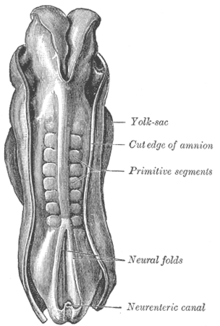
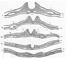
Following gastrulation, it emerges from the layer of cells known as the ectoderm (outer germ layer ) on the surface of the embryo , which is located above the notochord and, due to its influence, forms the slightly thicker neuroectoderm here as the neural plate . This bulges up laterally with neural bulges and in the middle between them to form an elongated neural groove . Then this neural groove folds down with the neural folds to form a tubular structure. Through this process, called primary neurulation , the neural tube is formed and sunk inwards and is separated from the ectoderm, which closes again as a surface ectoderm over the folded-over appendix of the central nervous system . On both sides along the neural tube form which temporarily the neural crest - from cells of the peripheral area of the former neural plate - from which a later date. a. In addition to melanocytes ( pigment cells ), various parts of the peripheral nervous system emerge.
From cells of the old center portion of the neural plate so the neural tube from which at forming vertebrates , the spinal cord and the brain develop. In lancet fish it is retained in its basic form and becomes the (central) nervous system. The larvae of salps also show a neural tube, but their ontogenetic development regresses.
In the human embryo, the neural tube is created between the 19th and 28th day of its development. The end to the neural tube begins from the middle, the area of the later hindbrain . The end of this development phase is the closure of the two neural tube openings, first the neuropore anterior (rostralis) in front with the lamina terminalis (about 29th day), then the neuropore posterior (caudalis) at the back (about 30th day). Disorders of development in this phase can lead to neural tube defects , and thus to spina bifida or anencephaly .
The lumen of the neural tube later becomes liquor-carrying cavities: the central canal of the spinal cord and the ventricular system of the brain with the aqueduct . Together they represent the inner CSF space .
Individual evidence
- ↑ Benninghoff: Macroscopic and microscopic anatomy of humans, Vol. 3. Nervous system, skin and sensory organs . Urban and Schwarzenberg, Munich 1985, ISBN 3-541-00264-6 , pp. 65ff.

