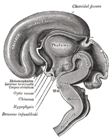
Lamina terminalis

The lamina terminalis (lat. Lamina 'layer', 'thin plate'; terminalis 'delimiting') is an unpaired thin tissue plate in the brain across the median plane . Located above the optic chiasm , it forms the anterior wall of the third cerebral ventricle and contains the organum vasculosum laminae terminalis . In embryonic development, the lamina terminalis represents the anterior closure of the forebrain after the cranial closure of the neural tube , up to the outgrowth of the two hemispheres of the endbrain , between which it then lies.

At the dorsal edge of the lamina terminalis, medial portions of both hemispherical vesicles merge to form the lamina reuniens . In this area the transversely connecting commissure systems of the telencephalon arise : the commissura anterior ( commissura rostralis ), the commissura fornicis ( commissura hippocampalis ) and finally the corpus callosum (bar).
Individual evidence
- ^ Anatomy Volume 2, Benninghoff - Drenckhahn, Elsevier Urban & Fischer, 16th edition 2004 ISBN 3-437-42350-9
- ↑ Benninghoff: Macroscopic and microscopic anatomy of humans, Vol. 3. Nervous system, skin and sensory organs . Urban and Schwarzenberg, Munich 1985, ISBN 3-541-00264-6 , p. 89 and p. 143, respectively.