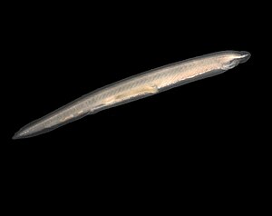
Lancet fish
| Lancet fish | ||||||||||||
|---|---|---|---|---|---|---|---|---|---|---|---|---|

Lancet fish Branchiostoma lanceolatum |
||||||||||||
| Systematics | ||||||||||||
|
||||||||||||
| Scientific name of the class | ||||||||||||
| Leptocardii | ||||||||||||
| Müller , 1845 | ||||||||||||
| Scientific name of the family | ||||||||||||
| Branchiostomatidae | ||||||||||||
| Bonaparte , 1846 |
The representatives of the Branchiostomatidae family from the order Amphioxiformes are referred to as lancet fish . As far as we know today, they are the only recent skullless . The lancet fish are often referred to as "Amphioxus" (from the Greek: "pointed on both sides"). Amphioxus is an outdated synonym of the genus Branchiostoma . It is used as a common name , especially in Anglo-Saxon usage, alongside lancelet .
The last common ancestors of lancetfish and vertebrates lived approximately 550 million years ago.
construction
The lancet fish are 4–7 cm long, unpigmented, sometimes iridescent organisms with a worm-like habit . The front end is characterized by a free-standing rostrum , the rear end by a small fin. Ventral to the rostrum is the mouth opening (14) , which is surrounded by long mouth cirrus (13). In the front area of the body (gill intestine region) the lancet fish are almost triangular in cross-section, the tail is flattened on the side. From the rostrum onwards there is an evenly high fin edge . There are no external attachments such as gills or paired fins.
As axial skeleton which serves notochord (2), which is characteristic of the phylum Chordata. It is a dorsally located, elastic axial rod between the spinal cord (1) and the intestine (11), which runs through the animals from the front to the rear end. The notochord is also covered by a coarse, connective tissue, notochord sheath. It serves as an endoskeleton and thus gives the Branchiostoma protection, strength and serves as a starting point for the muscles. The notochord arises ontogenetically from the mesoderm of the tricotyledonous deuterostomia (in which the original mouth develops into the anus).

Most of the musculature consists of striated muscle cells and is found segmentally arranged on the animal's flanks. Those myomers (muscle segments), separated by connective tissue myosepta , are put inside each other like a sugar bag and are braced by connective tissue with the chorda dorsalis and with each other, they are used for locomotion. Similar to the Kahn muscle cells of nematodes send the individual muscle cells in the myomeres cytoplasmic streamer for neural tube (3) in order to "pick up" their excitation there, i.e. the neuromuscular synapses lying at the side surfaces of the central nervous system and not, as in the skull animals , in Muscle itself. On the underside of the animal lies the also striated “wing muscle” ( musculus pterygoideus ), the contraction of which compresses the gill intestine (11) and the peribranchial space (18) and thus serves to “cough up” larger particles. Its innervation takes place in a “conventional” way, that is, through the branches of motor nerve cells whose cell bodies lie in the neural tube. Remarkably - and unlike all other chordates - the chorda dorsalis itself consists of striated muscle cells stacked one behind the other like a roll of money, which, like the muscles of the myomers, "pick up" their innervation from the central nervous system via cytoplasmic extensions. Smooth muscles are only sparsely present, they are mainly located in the walls of the coelom spaces .
The blood circulation is closed, the circulation is driven by contractile vascular sections, especially the gill intestine area. The blood passes from the sinus venosus into a cranially adjoining ventral aorta (endostyl artery ), from where it is passed on via the gill arch vessels to the dorsal (paired) aortic roots . The dorsal aortic roots unite in the ventral direction to form the dorsal aorta , which merges into the caudalaorta behind the anus opening. From the tail area, the blood reaches the subintestinal vein via the caudal vein , and from the muscle segments it reaches the cardinal veins. The cardinal veins flow into the sinus venosus on both sides via a ductus cuvieri each , the subintestinal vein initially receives the blood from the intestinal capillary network. The liver sac is supplied with venous blood rich in nutrients from the intestine via the portal vein . This travels from the capillary network of the liver blind sack into the hepatic vein and from there into the sinus venosus. This hepatic portal vein system is considered to be the synapomorphism of the skullless and cranial animals . The blood vessels of the lancet fish are not lined endothelially , the term veins and arteries is only used because of the direction of blood flow.
The secondary body cavity ( coelom ) is subdivided into sclerocoeles , narrow myocoeles , coelomas in the fin chambers, paired subchordal coelomas , endostylarcoelomas , gonadal coelomas , metapleural cavities and coelomal residues anterior to the transverse muscle ("wing muscle"). Coelom tubes running in the main gill arches connect the subchordal coelom with the endostylarcoelom.
The mouth opening is surrounded by mobile cirrus that carry chemoreceptors . The oral cavity (vestibule) is delimited laterally by cheeks and posteriorly by a tentacle-bearing velum . Eyelash fields sit on the cheeks, the wheel organs . In the dorsal roof of the mouth there is a depression with an eyelash flame, the Hatschek's pit . The pharynx , which lies freely behind the velum in the peribranchial space, is designed as a gill intestine , that is, it is pierced by gill slits. There are gill arches between the gill slits. Support rods and blood vessels run in the gill arches. The endostyle ( see chordata ), also known as the hypobranchial groove, is located in the center of the gill gut , and a ciliate groove, the epibranchial groove, on the dorsal side. Separated from the gill intestine by an iliocolon ring , the middle and rectum adjoin this. The lancet fish have a liver blind sack ( caecum ), which is homologated with the liver of the cranial animals due to its location and due to the presence of a portal vein system.
The central nervous system consists of a neural tube or spinal cord with a narrow central canal running through the animal dorsal to the notochord (3). This tube widens rostrally to form a small vesicle (1), which should not be homologated with the brain or brain sections of cranial animals. Segmental spinal nerves extend from the spinal cord . There are no eyes or large sense organs, but pigmented light sense organs are found scattered across the spinal cord. The animals can at least differentiate between light / dark and directions.
In Branchiostoma lanceolatum, food is consumed through the mouth opening (14) behind the cranial cranial cranium (13), the inflowing water is filtered by the gill intestine behind it for nutrients, which thus reach the digestive tract and the hepatic blind sac. Excrement is excreted through the anus (5). The absorbed water passes through the gill slits (11), which are surrounded by fine blood capillaries for oxygen absorption, into the peribranchial space (18), where it is first collected and then released through the atriopore . Breathing occurs not only through the gill slits, but to a large extent also through the skin.
The excretion takes place via cyrtopodocytes , they are a mixture of proto- and metanephridia and are therefore unique in the animal kingdom: extensions of cells of the coelom wall lie on the basal matrix of blood vessels. Ultrafiltration thus takes place on the wall of the primary body cavity (which is represented as the remainder of the blood vessel). The Coelom fluid corresponds functionally to the primary urine . This principle is implemented in all coelomat animals.
Way of life
The lancetfish are filter feeders . They filter plankton , detritus and other small organic particles from the surrounding sea water. When eating, lancet fish are usually buried in the ground, while the front end protrudes with the mouth wide open. A nutrient flow is generated by the eyelash flame of Hatschek's pit, the wheel organ and the cilia of the gill intestine. Water is trickled into the mouth opening, larger particles are prevented by the oral cirrus from entering the mouth opening. Another sorting takes place at the Velum. The nutrient flow reaches the gill intestine behind the velum. In the gill intestine, the endostyle creates a mucous film which is transported over the gill region to the opposite epibranchial groove. The nutrient water flow is filtered through this fine film of mucus. The water passes through the mucous film, between the gill arches and into the peribranchial space, from which it is excreted through the atriopore. The food particles, on the other hand, remain caught in the mucous film and are transported with it to the epibranchial groove. There, the slime film is rolled up into a feed sausage and transported into the subsequent intestine.
Lancet fish can move sideways by snake-like (undulating) movements. They are fast swimmers and can also move about in the ground to a limited extent. The lancet fish lie on their side on a hard substrate.
Reproduction and development
Lancets are separate sexes. The sex cells mature in gonads in myocoel areas along the side wall of the peribranchial space. The release occurs when the gonads burst open, and the sex products enter the peribranchial space and through the atriopore into the open water.
The lancet zygotes furrow radially and gastrulate by intussusception (invagination and invagination of the endoderm). Contrary to what is often shown, the neurulation does not take place, as in vertebrates, by rolling the neural tube, but rather the neural ectoderm is separated from the epidermal ectoderm and overgrown by the latter. Under the epidermal ectoderm, the neural ectoderm then rolls into the neural tube. An asymmetrical larva goes through a metamorphosis to adult.
Habitat and Distribution
Lancet fish occur in the sand or mud on the coasts of warm and temperate seas. Branchiostoma lanceolatum is native to the Black Sea or the Mediterranean, but also on the coasts of France, England and Scandinavia. Other species are also found in the Indian Ocean and Australia.
There are also occurrences on Helgoland , the Doggerbank and in the Kattegat , but not in the Baltic Sea.
Systematics
- Branchiostomidae Bonaparte family 1841
- Genus Asymmetron Andrews 1893
- Asymmetron lucayanum Andrews 1893
- Asymmetron maldivense (Forster Cooper 1903)
- Asymmetron inferum Nishikawa 2004
- Genus Epigonichthys Peters 1876
- Epigonichthys australis (Raff, 1912)
- Epigonichthys bassanus (Günther, 1884)
- Epigonichthys cingalense (Kirkaldy, 1894)
- Epigonichthys cultellus Peters, 1877
- Epigonichthys hectori (Benham, 1901)
- Epigonichthys lucayanum (Andrews, 1893)
- Epigonichthys maldivensis (Foster Cooper, 1903)
- Genus Branchiostoma OG Costa 1834
- Branchiostoma africae Hubbs in Monod, 1927
- Branchiostoma arabiae Webb, 1957
- Branchiostoma belcheri (Gray, 1847)
- Branchiostoma bennetti Boschung et Gunter, 1966
- Branchiostoma bermudae Hubbs, 1922
- Branchiostoma californiense Andrews, 1893
- Branchiostoma capense Gilchrist, 1902
- Branchiostoma caribaeum Sundevall, 1853
- Branchiostoma elongatum (Sundevall, 1852)
- Branchiostoma floridae Hubbs, 1922
- Branchiostoma gambiense Webb, 1958
- Branchiostoma indicum (Willey, 1901)
- Branchiostoma lanceolatum (Pallas, 1774)
- Branchiostoma leonense Webb, 1956
- Branchiostoma longirostrum Boschung, 1983
- Branchiostoma malayanum Webb, 1956
- Branchiostoma nigeriense Webb, 1955
- Branchiostoma platae Hubbs, 1922
- Branchiostoma senegalense Webb, 1955
- Branchiostoma tattersalli Hubbs, 1922
- Branchiostoma virginiae Hubbs, 1922
- Genus Asymmetron Andrews 1893
Numerous authors synonymized the genus Asymmetron with Epigonichthys , both of which have a striking feature, the asymmetrical formation of the gonads. However, later investigations have shown that this classification incorrectly reflects the relationships. A family Asymmetronidae, which used to be occasionally differentiated, is no longer considered to be justified today.
Individual evidence
- ^ Nicholas H. Putnam et al .: The amphioxus genome and the evolution of the chordate karyotype. Nature , Volume 453, 2008, pp. 1064-1071; doi : 10.1038 / nature06967
- ↑ Teruaki Nishikawa (2004): A New Deep-water Lancelet (Cephalochordata) from off Cape Nomamisaki, SW Japan, with a Proposal of the Revised System Recovering the Genus Asymmetron. Zoological Science 21: 1131-1136.
Web links
- Branchiostoma lanceolatum or the lancet - accessed January 31, 2008
- Videos on Branchiostoma lanceolatum published by the Institute for Scientific Film .