
Tear sac

The lacrimal sac ( lacrimal sac ) is a part of the lacrimal apparatus . It is the upper widened part of the lacrimal duct and lies in a notch in the lacrimal bone and the frontal process of the upper jaw . It connects the lacrimal tubules, which drain the tears from the surface of the eyeball, and the lacrimal duct, which carries the fluid into the nasal cavity.
anatomy
The lacrimal sac is located in a bony depression of the lacrimal bone ( Fossa sacci lacrimalis ) at the corner of the eye on the side of the nose . This pit is bounded behind by the crista lacrimalis posterior of the lacrimal bone and in front by the crista lacrimalis anterior of the frontal process of the upper jaw .
The tear sac has an oval shape. In humans, it has a length between 12 and 15 millimeters and a volume of 20 to 100 mm 3 . The upper end is blindly closed, which is called the lacrimal sac vault ( Fornix sacci lacrimalis ). The tear ducts open individually or together below the vault into the tear sac. The lower end tapers into the lacrimal duct.
The wall of the lacrimal sac is fused with the periosteum of the lacrimal bone and with the periorbita , so that its lumen is always kept open. The anterior surface is covered by the inner lid ligament ( ligamentum palpebrale mediale ). The posterior surface is crossed by the lacrimal part of the orbicularis oculi muscle , which is attached to the lacrimal bone.
Like the lacrimal duct, the tear sac is lined by a two-row, highly prismatic epithelium.
Function and clinical significance
The sack is mainly used to drain the surface of the eyeball when there are large amounts of tear fluid and also helps to remove dirt, bacteria and other foreign bodies from it. The continuous flow of fluid is ensured by contractions of the orbicularis oculi muscle , which has a pumping function on the lacrimal sac.
Inflammation of the bags under the eyes is known as dacryocystitis .
The tear sac can be imaged using dacryocystography . Here, contrast agents introduced and x-rays taken of it.
Additional pictures

nasal wall of the left eye socket

the left orbicularis oculi muscle seen from behind

the lacrimal system on the right side (the lacrimal sac is visible on the top right)
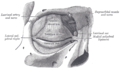
the tarsi and their ligaments on the right front side (the tear sac can be seen in the middle right)


Web links
Individual evidence
- ^ A b Hanson Kelly Corning: Textbook of topographical anatomy for students and doctors . Springer-Verlag, 20th edition 2013, ISBN 978-3-662-29954-8 , p. 67.
- ^ Albert J. Augustin: Ophthalmology . Springer-Verlag, 2nd edition 2013, ISBN 978-3-662-05919-7 , p. 1140.
- ^ Richard Funk et al .: Pocket Textbook Anatomy . Georg Thieme Verlag, 2010, ISBN 978-3-13-162511-3 , p. 663.
- ^ Astrid Kruse Gujer, Christine Jacobsen, Klaus W. Grätz: Specialist knowledge of oral and maxillofacial surgery . Springer-Verlag, 2013, ISBN 978-3-642-30003-5 , p. 8.
- ↑ a b L.C. Junqueira, J. Carneiro: Histology: cytology, histology and microscopic human anatomy. Taking into account the histophysiology . Springer-Verlag, 4th edition 2013, ISBN 978-3-662-07780-1 , p. 681.