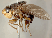
Tumble Fly
| Tumble Fly | ||||||||||||
|---|---|---|---|---|---|---|---|---|---|---|---|---|
| Systematics | ||||||||||||
|
||||||||||||
| Scientific name | ||||||||||||
| Cordylobia anthropophaga | ||||||||||||
| ( Blanchard , 1872) |
The tumbu fly ( Cordylobia anthropophaga ), also often called the mango fly, is an ectoparasite from the genus Cordylobiae and thus belongs to the family of blowflies (Calliphoridae).
It occurs in tropical Africa south of the Sahara and lays its eggs mainly on sandy soils.
Tumbu fly (also mango fly) as a cause of disease
After hatching, the larvae can penetrate the skin of the person through direct body contact or through laundry left on the floor to dry, for example, after the person has put on the clothing again. They then mature in the subcutaneous tissue within about two weeks . In doing so, they cause dermatomyiasis (infection of the skin with fly larvae; myiasis = "maggot damage").
Diagnosis of myiasis caused by tumbu flies
A small number of returnees from the tropics have infectious skin changes ( dermatomyiosis ), which z. B. caused by the Tumbu fly. Practical as well as clinical doctors sometimes have problems clearly diagnosing this disease, since it resembles an inflammation of the hair follicles in the initial stages, as in acne vulgaris . However, this follicle can develop into a walnut-sized bump in a period of 8-10 days, before the maggot leaves the skin to pupate. When magnifying the magnifying glass, a central blackish point can often be seen, which corresponds to the anus and respiratory organ of the maggot.
The largest reservoir for the reproduction and distribution of blowflies are usually animals, but the genus Cordylobia anthropopha also likes to attack humans.
Course of disease
Within a few days, a firm, elastic, but less painful subcutaneous swelling with a diameter of about one to three centimeters forms around the larva. In the middle of the swelling there is a central opening that allows the maggots to breathe and from which serious, bloody secretions are sometimes emptied with fine vesicles. As the larva grows, the itchy, painful movements of the larva can be noticed. The finished third larvae of the fly hatch from the same opening after about eight to twelve days.
Lymph node swelling, fever are rare, secondary infections can develop if the larva is improperly removed or if remnants of the larval body (e.g. mouth tool) remain in the follicle when attempting to remove it.
therapy
As long as they are still small, the larvae can sometimes be expressed like a so-called "blackhead". In the advanced stage, the whitish larval body becomes visible by simply widening the central breathing opening with a sharp cannula (e.g. gauge 20) and can then be carefully grasped with fine anatomical tweezers and pulled out completely with constant tension. The completeness of the larva must be checked, which then regularly tries to free itself from the tweezers. A bloodless, usually successful method consists of closing the breathing opening with petroleum jelly or oil. This forces the maggots to breathe out of the skin cavity. Furthermore, you can attract the maggots with slices of bacon placed on the swelling. As a rule, the maggots will attach themselves to the bacon slices over the course of one to two hours and can then be easily removed afterwards.
Prevention and avoidance
The transfer of eggs to the body usually takes place via clothing or bed linen, if they were not completely dry in a warm, humid climate and / or were contaminated with faeces / urine. Laundry should therefore always be exposed to intense sunlight until it is straw dry. Diapers in particular should be ironed whenever possible. Particular care should be taken with waistbands and clothing parts that contain double layers of textile and therefore dry later. Cold treatment in the freezer below −20 ° Celsius does not safely kill the larvae. The skin changes are therefore mainly found on skin that is covered by clothing, such as the torso and legs. Thorough examination of the skin of the entire body, especially the genital and anal area, is recommended, as several lesions (damage, injury or disruption of body tissue) with several larvae per wound are possible.
source
- Albrecht von Schrader-Beielstein: Differential diagnosis in skin diseases. In: Rüdiger Braun (Hrsg.): Travel and Tropical Medicine: Course book for further training, practice and advice. Schattauer, Stuttgart / New York 2005, ISBN 3-7945-2286-9 , p. 154.
Individual evidence
- ↑ EGNauck: Textbook of tropical diseases . Ed .: Werner Mohr, Hans-Harald Schuhmacher, Fritz Weyer. Georg Thieme Verlag, Stuttgart 1975, ISBN 3-13-380704-8 , p. 26-28 .
- ^ Paul S. Auerbach et al .: Field Guide to Wilderness Medicine . 4th edition. Elsevier, Mosby, ISBN 978-0-323-10045-8 , pp. 459-460 .
- ↑ Dr. Jane Wilson-Howarth: Bugs Bites & Bowels . Ed .: CadoganGuides. 2013, ISBN 978-1-86011-332-1 , pp. 271-272 .

