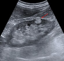
Angiomyolipoma
| Classification according to ICD-10 | |
|---|---|
| D30 | Benign neoplasm of the urinary organs |
| D30.0 | - kidney |
| ICD-10 online (WHO version 2019) | |
When angiomyolipoma is a rare, benign tumor of the kidney that a high proportion of fat tissue has.
They mostly occur in women between the ages of 40 and 60. 80% of the cases are asymptomatic angiomyolipomas in both kidneys, which means that they do not cause any symptoms. There is an equal distribution between men and women in these patients. The maximum age in this group of patients is 30 years of age. In some of the patients angiomyolipoma is associated with tuberous sclerosis (Bourneville-Pringle's disease).
pathology

Angiomyolipoma originates from the perivascular epithelial cells , the growth of which is probably controlled in a hormone-dependent manner . The angiomyolipoma does not metastasize, but in 30% of cases it can grow into the fatty tissue surrounding the kidney, renal pelvis and even into the renal veins. The tumor accounts for 1% of all surgical findings. Angiomyolipoma is 20% associated with tuberous sclerosis.
Macroscopically, these are unencapsulated gray-yellow lesions that can be round to oval. The angiomyolipoma partially shows a multicentric growth. Occasionally, lymph node involvement can occur.
Mature fat cells and smooth muscle cells can be detected microscopically. The muscle cells can show numerous mitoses , so the tumor can be misinterpreted as a sarcoma . In addition, there are atypical blood vessels.
Diagnosis and therapy


The symptoms are unspecific, flank pain may occur. Therefore, imaging tests can provide a possible indication of the diagnosis (see below). A potentially life-threatening complication of angiomyolipoma is bleeding through spontaneous rupture into the retroperitoneum ( Wunderlich syndrome) . Pregnancy can increase the risk of rupture.
diagnosis
Imaging procedures:
- In the ultrasound there are strong echogenic masses in the kidney due to the fat content.
- The representation of the blood vessels is not helpful for the differential diagnosis, since in angiomyolipoma, as in renal cell carcinoma, new vascular formations occur.
- In the computed tomography A contingent of the high fat content hypodense mass shows in the kidney with -20 to -80 units Hounsfield . This enables a differentiation from renal cell carcinoma . Calcifications do not occur in angiomyolipomas.
- In the case of MRI , evidence of a high fat content suggests an angiomyolipoma and not renal cell carcinoma.
therapy
When the angiomyolipoma than 4 cm is greater, with strong symptoms or with tumors of unknown dignity a partial removal (resection) of the kidney should be sought.
Selective embolization is a possible minimally invasive therapy option. With this technique, however, recurrences can occur and drainage of the necrotic tissue may be necessary.
swell
literature
- Riede / Schäfer Pathology ISBN 3-13-683303-1
Web links
- Macroscopic image of an angiomyolipoma on PathoPic
- Microscopic image of an angiomyolipoma stained with HE on PathoPic
- Sonographic images of angiomyolipoma