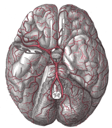
Anterior cerebral artery
The anterior cerebral artery ( Latin: anterior cerebral artery ), known in animals as the rostral cerebral artery , is one of the three main arterial vessels of the brain . It rises in the area of the circle of Willis at the base of the brain from the internal carotid artery . Before the optic nerve junction , it forms an anastomosis with the opposite side, called the anterior communicating artery . This precommunicative section is called the A1 segment. The postcommunicative segment, the A2 segment, runs between the cerebral hemispheres around the bar . The anterior cerebral artery is also referred to here as the pericallosal artery .
Coverage area
The coverage area is subject to a certain individual variability. As a rule, this includes the anterior parts of the hypothalamus , most of the basal ganglia (via the central arteries ), the basal surface of the frontal lobe and the medial hemispherical surface of the frontal and parietal lobe up to approx. 1 cm above the mantle edge . In the end zones of its supply area it forms small anastomoses with the arteria cerebri media and the arteria cerebri posterior . Short branches supply the optic chiasm , the optic nerve and the optic tract .
Failure symptoms
An occlusion of the anterior cerebral artery leads to paresis with a pronounced leg and variable impairment of motor function through damage to the supplementary motor cortex . If the Aa. centrales are involved, lesions of the internal capsule can result in aphasia , contralateral hemiparesis and central facial paresis .
Web links
Individual evidence
- ↑ Thomas Deller: Sobotta textbook anatomy . Ed .: Jens Waschke, Tobias M. Böckers, Friedrich Paulsen. Urban & Fischer Verlag / Elsevier, Munich 2015, ISBN 978-3-437-44080-9 , 11.5.6 Topography and supply areas of the arteries.

