Atheroma
| Classification according to ICD-10 | |
|---|---|
| L72.1 | Trichilemmal cyst |
| ICD-10 online (WHO version 2019) | |
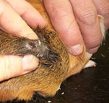
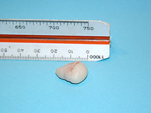
An atheroma ( Attic Greek for "wheat groats ") or a trichilemmal cyst ( synonym Tricholemmalzyste ), in German sebum cyst , is a pinhead to chicken egg-sized, rarely to apple-sized, benign cyst in the subcutaneous tissue . In the vernacular , these cysts are also known as cheesefeet , Balggeschwulst or semolina node designated.
Emergence
Atheromas usually arise as a result of a blockage of the duct for the sebum secretion . They consist of fat droplets , fat crystals and epidermal cells. Atheromas are found individually or in large numbers, mostly on the hairy head , on the face and neck , between the abdomen and neck, but also in other places (e.g. in the genital area and the chest).
A distinction is made between real and false atheromas. Real atheromas are epidermoid cysts, consist of embryonically scattered epidermis or gland cells, have no opening and are usually located on the hairy scalp, on the forehead or around the eye. False atheromas are sebum retention cysts, i.e. they consist of blocked gland secretions from the hair sebum glands , have an opening and are usually found on the face, neck, chest, back and scrotum .
therapy
Discomfort occurs only when the atheroma becomes inflamed or infected with purulent disease; it is then most conveniently removed surgically. It should be noted, however, that removing an infected atheroma is problematic, as there is a risk that the bacteria will spread throughout the body during the operation. So, first of all, it is necessary to remove the contents of the atheroma. After that, the sack can refill with fat and a new cyst can develop at the operated site, but experience has shown that this is not the case for a long time. The complete removal can be done with renewed swelling and before infection, or after infection under anesthesia.
Atheromas can grow in size at different rates. They are surgically removed in the event of inflammation, rapid increase in size, preventive or for cosmetic reasons, usually under local anesthesia.
literature
- H. Sharma, A. Sinha, B. J. Singh: Sebaceous cyst presenting with necrotizing ulcerative infection over trochanteric area mimicking necrotizing fasciitis. in: J Eur Acad Dermatol Venereol . 2006; 20 (3): 345-346. doi : 10.1111 / j.1468-3083.2006.01405.x
- B. Kanee: Removal of Sebaceous Cysts. A Modification in Technique. In: Can Med Assoc J . 1963; 89 (10): 518-519. PMC 1921887 (free full text)
See also
Web links
Individual evidence
- ^ Pschyrembel Clinical Dictionary, de Gruyter, 1977.


