Heart skeleton

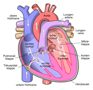
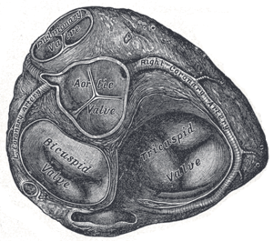
The heart skeleton is a layer of connective tissue , often with fat , cartilage or bone deposits , which separates the atria and the chambers of the heart just above the valve level . The heart skeleton includes four fiber rings ( anuli fibrosi ) to which the heart valves are attached by connective tissue. These fiber rings are
- the right fibrous ring ( annulus fibrosus dexter ) around the tricuspid valve
- the Pulmonalisring ( annulus pulmonary trunk ) to the pulmonary
- the left fibrous ring ( annulus fibrosus sinister ) around the bicuspid valve or mitral valve
- the aortic ring ( anulus aortae ) around the aortic valve
The heart skeleton is used to mechanically stabilize the openings of the heart chambers and the origin and insertion of the heart muscles . It separates the cardiac muscles of the atria from those of the ventricles and thus enables them to contract at different times. In addition, the cardiac skeleton seals off the conduction of excitation from the atria, so it serves as electrical insulation. The excitation resulting from the stimulus from the atria can therefore only be passed on into the ventricles via the bundle of His , which pierces the dextronum fibrosum.
The heart skeleton (central valve level) is crucial for the mechanics of the heart's action: Due to the recoil during the expulsion of blood, the apex of the heart is relatively locally fixed over the entire heart cycle and hardly moves. Consequently, when the ventricular muscles contract, the valve plane is pulled downwards in the direction of the apex of the heart. There is a pull on the large heart vessels, which now contract reflexively when the ventricular muscles relax and thus pull the valve level up again. The mechanics of the heart is therefore a “raising and lowering of the valve level”. Chamber muscles and heart vessels work in opposite directions.
literature
- Uwe Gille: Cardiovascular and immune system, Angiologia. In: Franz-Viktor Salomon, Hans Geyer, Uwe Gille (Ed.): Anatomy for veterinary medicine. Enke, Stuttgart 2004, ISBN 3-8304-1007-7 , pp. 404-463.

