Heterochromatin
| Parent |
| Chromatin |
| Gene Ontology |
|---|
| QuickGO |
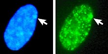
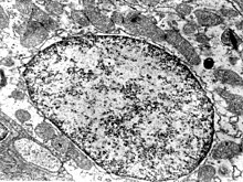
As heterochromatin compressed is chromatin in the cell nucleus called, which can be stained well. Here the deoxyribonucleic acid (DNA) is strongly bound to histone and non- histone proteins. This means that the genetic information remains largely inactive. The term heterochromatin is used to distinguish it from euchromatin , which is more easily accessible. Heterochromatin is colored more strongly (positively heteropyknotic ) than euchromatin by various light microscopy staining techniques (e.g. Feulgen reaction , Giemsa staining ) . In the transmission electron microscope , too, it appears dark because of its strong electron absorption.
Types of heterochromatin

- Constitutive heterochromatin is often close to the centromeres and is then also called centromeric heterochromatin . Numerous copies of repetitive DNA lie directly behind one another , but no genes. Constitutive heterochromatin is formed in all cells of an organism on the same sections of the chromosomes, hence the name.
- In contrast, facultative heterochromatin is only formed in a few cells of an organism. The best-known example is the Barr body , one of the two X chromosomes in the cells of female mammals . It is shut down in order to match the gene dose of a female mammal with a male X, Y individual, and thereby becomes heterochromatic. In female organisms with an extra X chromosome ( trisomy X ), two X chromosomes are heterochromatically shut down, so that there are often hardly any symptoms.
The classic primary marker protein for heterochromatin is the highly conserved heterochromatin protein 1 (HP 1). Another characteristic feature of heterochromatic regions are hypoacetylation (the absence of acetyl groups) and methylation on certain amino acids of the histones.
Aggregates of heterochromatin are called chromocentres .
history
Using the technical terms heterochromatin and euchromatin , Emil Heitz specified the word chromatin proposed by Walther Flemming : the non-heterochromatic chromatin areas are euchromatic. In addition, Heitz observed for the first time in Drosophila virilis that in endoreplicating cell nuclei the (constitutive) heterochromatin multiplies far less than the euchromatin or not at all.
Web links
- Medical Dictionary: heterochromatin (English)
Individual evidence
- ^ Emil Heitz: Heterochromatin, Chromocentren, Chromomere. In: Reports of the German Botanical Society. 47/1929, p. 277.
- ↑ Walther Flemming: Contributions to the knowledge of the cell and its life phenomena. Part II. In: Archive for Microscopic Anatomy 18: 151-259, 1880. There p. 158.
- ^ Emil Heitz: About α- and β-heterochromatin as well as constancy and structure of the chromomers in Drosophila. In: Biologisches Zentralblatt 54: 588-609, 1934. There p. 596.
