
Bone marrow edema
Bone marrow edema is a term used in medical imaging using magnetic resonance imaging (MRI). It stands for edema-equivalent signal changes d. H. increased signal intensity (light) in T2-weighted sequences and decreased signal intensity (dark) in T1-weighted sequences in cancellous bone structures. The signal intensity can be measured in grayscale (necessarily as a proportion of a reference structure, e.g. muscle, shown with it). Bone marrow edema cannot be shown by X-rays ( X-ray , computed tomography ).
Assignment of the signal changes to changes in the bone
The changes in brightness in the MRI are based on locally increased intravascular or extravascular fluid ( blood serum , tissue water, lymph , hypervascularization ) with various pathological changes: injury-related, tumor-related, infection-related, rheumatic (inflammatory processes with locally increased new formation of particularly permeable capillaries), but inflammatory also in the case of reparation processes with the formation of new granulation tissue / callus (for the temporary closure of an injury-related bone defect, connective tissue neoplasia). Bone marrow edema sites show radionuclide build-up on bone scintigraphy . Bone marrow edema due to microfractures limited to the cancellous bone - without accompanying cortical damage - can be painless. In the acute state of a bone injury - in addition to the edema-equivalent signal changes in the bone - these are always observed in the surrounding soft tissues. The transition from the acute injury-related inflammatory, painful bone marrow edema to the healing-related painless bone marrow edema cannot be demarcated morphologically; the regression of the edema-equivalent signal changes in the soft tissues and the decrease in pain symptoms can provide corresponding indications. The regression of traumatic bone marrow edema under therapy corresponds to the course of fracture healing. Normal blood-forming bone marrow, e.g. B. in the vertebral bodies of young people, appears relatively bright in T2-weighted sequences (and relatively dark in T1-weighted sequences) compared to the normal fatty bone marrow in the vertebral bodies of older people.
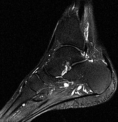
- Foot, MRI, T2 weighting

Bone marrow edema (light) in the talus, scaphoid and sphenoid bone
For comparison: ankle, scaphoid and sphenoid bone are normal


- Foot, x-ray

Existing bone marrow edema not recognizable

Historical
The term bone marrow edema (bone marrow edema) coined in 1988 AJ Wilson and staff. They found local hyperemia, increased bone metabolism and radionuclide accumulation (in the bone scintigram) in areas with decreased signal intensity in T1-weighted sequences and increased signal intensity in T2-weighted sequences. They suspected local water retention in the bone marrow to be the cause.
literature
- AJ Wilson, WA Murphy, DC Hardy, WG Totty: Transient osteoporosis: transient bone marrow edema? In: Radiology . No. 167 , 1988, pp. 757-760 , doi : 10.1148 / radiology.167.3.3363136 .
- Johan L Bloem, Monique Reijnierse, Tom WJ Huizinga, Annette HM van der Helm-van Mil: MR signal intensity: staying on the bright side in MR image interpretation. In: RMD Open. 4, 2018, p. E000728, doi : 10.1136 / rmdopen-2018-000728 .
- R. Wunsch: What should the pediatrician know about magnetic resonance imaging? In: Monthly Pediatrics . tape 150 , no. 12 , 2002, p. 1523-1533 , doi : 10.1007 / s00112-002-0626-5 .
Individual evidence
- ^ HE Daldrup-Link, T. Henning, TM Link: MR imaging of therapy-induced changes of bone marrow. In: European radiology. Volume 17, number 3, March 2007, pp. 743-761, doi : 10.1007 / s00330-006-0404-1 , PMID 17021706 , PMC 1797072 (free full text) (review).
- ↑ Suzanne S. Long, Corrie M. Yablon, Ronald L. Eisenberg: Bone Marrow Signal Alteration in the Spine and Sacrum . In: American Journal of Roentgenology . tape 195 , September 2010, p. W178 , doi : 10.2214 / AJR.09.4134 (English).
- ↑ rsna.org: Transient osteoporosis: transient bone marrow edema? published June 1, 1988, loaded July 23, 2019