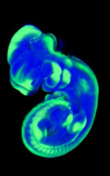
Optical projection tomography
The optical projection tomography (OPT) is an imaging process of medical diagnosis and biochemical analysis. It represents slightly or highly transparent objects, such as cell tissue, in a spatial manner and uses visible light for this. Optical projection tomography is regarded as the X-ray-free counterpart to computed tomography and is also known as "CT with light". In biochemistry, the process is part of optical microscopy in the broadest sense .
Mode of action
OPT works with relatively small, commercially available devices and uses open source software to evaluate the images. In principle, one shines through the tissue from different angles with visible light and collects the reflected rays according to direction and frequency. From this "filtered reverse transformation" one can then produce a volume model of the tissue from slice images using methods known from CT and make structures such as cell changes or tumors visible, which would otherwise only be possible by actually cutting (sectioning) the tissue. As a rule, the examined material must first be made sensitive to certain light components, for example by adding fluorescent substances.
Optical projection tomography has only played a role in research since the early 2000s because of the high computational effort and has gradually been establishing itself in the practice of biochemistry and medicine since the 2010s. It competes with several optical imaging processes, such as photoacoustics , where a pulsed laser is used to bombard tissue and the acoustic reflections are measured.
Web links
- OPT - 3D visualization with visible light ( Memento from April 13, 2016 in the Internet Archive ) Laser Center of the University of Hanover
- Device for optical projection tomography with a rotatable sample holder for imaging a sample . Patent DE 60223728 T2, by Paul Ernest Perry and James Alexander Sharpe. Entry in Google Patents
- Novel tomographic imaging method ( Memento from April 13, 2016 in the Internet Archive ) In: Biophotonik, May 2013
