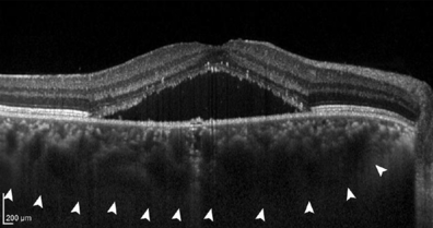
Macular pachychoroid disorders
Under pachychoroidal diseases of the macula one understands a group of diseases, especially of the retina ( retina ) of the eye , in which the choroid ( choroidea ) below the retina is pathologically thickened and congested. As a result, the surrounding structures damaged, particularly the retina (retinal) , and the retinal pigment epithelium . This leads to impaired vision. The best-known representative of the spectrum of pachychoroid diseases, retinopathia centralis serosa , is the fourth most common cause of irreversible damage to the visual pit ( yellow spot , macula).
The term "pachychoroid" was first introduced in 2013 by David Warrow, Quan Hoang and K. Bailey Freund.
Cause of illness
Due to the reasons that have not yet been fully clarified, there is an excessive increase in thickness of the choroid ( choroid ), usually directly below the yellow spot (macula), the visual center of the retina. It is assumed that the thickening of the choroid is due to a congestion of the numerous blood vessels running through it.
The choroid membrane, which is too thick and congested, leads to pressure damage, leakage of fluid from the vascular system and an insufficient supply of oxygen and nutrients. This results in damage to the retinal pigment epithelium and the retina ( retina ), which ensures that the central vision in the central retina (yellow spot , macula) deteriorates.
to form
Pachychoroid without disease value
- If there is only one choroidal thickening, usually over 350 or 300 µm without damage to the surrounding structures, one speaks of an uncomplicated pachychoroid . It is believed that a large proportion of the population has a thickened choroid with no other symptoms. These include above all young and far-sighted people.
Pachychoroid with disease value
Within the pathological pachychoroidal spectrum, diseases of the center of the retina (yellow spot , macula) are most common.
- If there is pressure damage to the fine blood vessels ( capillaries ) due to the continuous congestion in the blood vessel system of the choroid and a continuous leakage of fluid in the direction of the adjacent Bruch's membrane and the retinal pigment epithelium , the resulting damage in the pigment epithelium is called pachychoroidal pigment epitheliopathy (PPE). . Patients with PPE usually have no symptoms.
- If further damage to the pigment epithelium leads to a breakthrough of fluid under the retina, a chorioretinopathia centralis serosa (CRCS) occurs . At this stage, patients often see blurred and report a reduction in central visual acuity with the perception of a central “gray spot”. In the majority of patients, the fluid disappears within a few months, but occurs again in up to 50% ( relapse ). In some patients the fluid persists chronically; therapy with tablets or various laser methods is possible.
- In around 25% of all patients with a chronic course, after an average of 17 years, blood vessels grow from the choroid in the direction of the retina, a so-called choroidal neovascularization . This defines pachychoroidal neovasculopathy (PNV) , which can cause severe visual impairment through bleeding and scarring of the visual pit (macula). The anti-VEGF therapy, which is injected directly into the eye ( intravitreal ), has proven to be an effective therapy .
- If aneurysms develop within these CNV, one speaks of a pachychoroidal aneurysmal type 1 CNV (PAT1), previously also called polypoidal choroidal vasculopathy (PCV).
- In addition, a focal choroidal excavation can occur in all stages , which probably represents a scar retraction in the choroidal tissue.
Pachychoroidal diseases outside the center of the retina have also been described around the optic nerve .
- If the increase in choroidal thickness is particularly evident around the optic nerve , it can lead to swelling of the optic nerve and accumulation of fluid in the retina, which is known as the peripapillary pachychoroid .
classification
A classification has been proposed, but there are no data that plausibly demonstrate the gradual sequence of diseases.
| Pachychoroid diseases of the macula (after Siedlecki et al.) | |
|---|---|
| 0 | Uncomplicated pachychoroid (UCP) |
| I. | Pachychoroidal pigment epitheliopathy (PPE) |
| II | Central serous chorioretinopathy (CRCS) |
| III | Pachychoroid Neovasculopathy (PNV) |
| a) with neurosensory detachment (= with fluid under the retina) | |
| b) without neurosensory detachment (without fluid) | |
| IV |
Pachychoroidal aneurysmal type 1 CNV (PAT1)
(formerly: Polypoidal Choroidal Vasculopathy, PCV) |
Individual evidence
- ↑ Sezen Akkaya: Spectrum of pachychoroid diseases . In: International Ophthalmology . tape 38 , no. 5 , October 2018, ISSN 0165-5701 , p. 2239–2246 , doi : 10.1007 / s10792-017-0666-4 ( springer.com [accessed March 17, 2020]).
- ↑ a b c Chui Ming Gemmy Cheung, Won Ki Lee, Hideki Koizumi, Kunal Dansingani, Timothy YY Lai: Pachychoroid disease . In: Eye . tape 33 , no. 1 , January 2019, ISSN 0950-222X , p. 14–33 , doi : 10.1038 / s41433-018-0158-4 , PMID 29995841 , PMC 6328576 (free full text) - ( nature.com [accessed March 17, 2020]).
- ↑ a b David J. Warrow, Quan Hoang V., K. Bailey friend PACHYCHOROID PIGMENT EPITHELIOPATHY: . In: Retina . tape 33 , no. 8 , September 2013, ISSN 0275-004X , p. 1659–1672 , doi : 10.1097 / IAE.0b013e3182953df4 ( lww.com [accessed March 17, 2020]).
- ↑ Claudine E. Pang, K. Bailey friend PACHYCHOROID NEOVASCULOPATHY: . In: Retina . tape 35 , no. 1 , January 2015, ISSN 0275-004X , p. 1-9 , doi : 10.1097 / IAE.0000000000000331 ( lww.com [accessed March 17, 2020]).
- ↑ Kunal K Dansingani, Orly Gal-Or, Srinivas R Sadda, Lawrence A Yannuzzi, K Bailey Freund: Understanding aneurysmal type 1 neovascularization (polypoidal choroidal vasculopathy): a lesson in the taxonomy of 'expanded spectra' - a review: Aneurysmal type 1 neovascularization . In: Clinical & Experimental Ophthalmology . tape 46 , no. 2 , March 2018, p. 189–200 , doi : 10.1111 / ceo.13114 , PMID 29178419 , PMC 5900982 (free full text) - ( wiley.com [accessed March 17, 2020]).
- ↑ Hyewon Chung, Suk Ho Byeon, K. Bailey Freund: FOCAL CHOROIDAL EXCAVATION AND ITS ASSOCIATION WITH PACHYCHOROID SPECTRUM DISORDERS: A Review of the Literature and Multimodal Imaging Findings . In: Retina . tape 37 , no. 2 , February 2017, ISSN 0275-004X , p. 199–221 , doi : 10.1097 / IAE.0000000000001345 ( lww.com [accessed March 17, 2020]).
- ↑ Nopasak Phasukkijwatana, K. Bailey friend, Rosa Dolz-Marco, Mayss Al-Sheikh, Pearse A. Keane: PERIPAPILLARY PACHYCHOROID SYNDROME: . In: Retina . tape 38 , no. September 9 , 2018, ISSN 0275-004X , p. 1652–1667 , doi : 10.1097 / IAE.0000000000001907 ( ovid.com [accessed March 17, 2020]).
- ^ Jakob Siedlecki, Benedikt Schworm, Siegfried G. Priglinger: The Pachychoroid Disease Spectrum — and the Need for a Uniform Classification System . In: Ophthalmology Retina . tape 3 , no. December 12 , 2019, p. 1013-1015 , doi : 10.1016 / j.oret.2019.08.002 ( elsevier.com [accessed March 17, 2020]).
