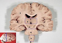
Pons
The pons ( Latin for "bridge") is a section of the brain that, together with the cerebellum, belongs to the metencephalon (rear brain).
Even at a cursory glance, the bridge is noticeable as a clearly raised transverse bulge that lies between the mesencephalon (midbrain) and the myelencephalon (medullary brain). Together with these, it forms the brain stem of the brain in the central nervous system .
anatomy

The bridge is divided into a front part, the base ( pars basilaris pontis ), and a rear part, the bridge hood ( tegmentum pontis or pars dorsalis pontis ). In animals, these are on top or bottom, depending on their body orientation.
Two longitudinal bulges can be seen at the base, through which the pyramidal tract ( Tractus pyramidalis = Tractus corticospinalis ) runs on each side . An important inflow for the blood supply to the brain, the basilar artery, runs in the central groove between the sulcus basilaris . The connecting line between the two halves, visible in cross-section, is crossed by numerous nerve fibers and is also called raphe ("seam"). Behind the transverse fibers of the pontine base lies the corpus trapezoideum (trapezoidal body), which represents the main intersection of the auditory pathway .

At the caudal edge of the bridge, the cranial nerves VII facial nerve and VIII vestibulocochlear nerve come to the surface of the brain dorsally in the cerebellopontine angle . In the sulcus bulbopontinus at the caudal base of the bridge, the VI. Cranial nerve abducens nerve den pons. The very strong V cranial nerve trigeminal nerve exits or enters laterally at the bridge.
The dorsal end of the bridge hood forms part of the floor of the rhomboid fossa ( fossa rhomboidea ) and thus the 4th ventricle . The connection to the cerebellum is established on both sides by the middle cerebellar limb ( Pedunculus cerebelli medius ).
tasks

On the one hand, the bridge is a passage area for all pathways that connect areas of the central nervous system in front of and behind . These include both ascending tracts, such as the lemnisci mediales , and descending tracts, such as the corticospinal tract , which establish connections between the cerebral cortex and the spinal cord .
The white matter of the pons contains, besides such longitudinal fiber strands ( Fibrae pontis longitudinales ), strong strands of fibers running across them ( Fibrae pontis transversae ). This also includes fibers crossing the midline ( raphe ) as well as both parts of the metencephalon, the pons with the cerebellum and the connecting fibrae pontocerebellares . A number of different bridging nuclei ( nuclei pontis ) are created for this, each of which represents switching stations, via which areas of the cortex in particular in the cortex of the cerebrum are connected to one another - mostly crossed - with those of the cerebellum . These nuclei of the bridge , which mediate projections of the cerebral cortex and the (contralateral) cerebellar cortex , are developed to a great extent in mammals , so that this bridge in the narrower sense only arises in them.
In the bridge hood, embedded in the pontine formatio reticularis , among other things, the motor nuclei of some cranial nerves are located, namely the nucleus motorius nervi trigemini of the V and the nucleus nervi abducentis of the VI. and nucleus nervi facialis of the VII.

