Metencephalon

- First three cerebral vesicles arise from the anterior neural tube ( prosencephalon , mesencephalon , rhombencephalon ; shown on the left half of the picture). - then the rhombencephalon differentiates into metencephalon and myelencephalon (right half of the picture). The metencephalon then differentiates further with the development of the cerebellum from the rostral diamond lips of the wing plate (not illustrated).
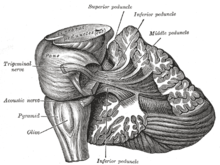
The metencephalon or hindbrain is part of the rhombencephalon ( hindbrain ) and consists of the bridge ( pons ) and the cerebellum (cerebellum).
The pons is divided into the bridge base ( pars basilaris pontis ) and the bridge hood ( tegmentum pontis ) attached to it . This extends to the diamond pit , the floor of the fourth cerebral ventricle ( ventriculus quartus ), which is bounded by the cerebellum towards the rear (or above).
The cerebellum closes dorsally (on the back) at the pons. It can also be viewed as the roof ( tectum ) of the metencephalon. The gray matter of the cerebellum is divided internally into core areas and externally into regions of its cortex ( cortex cerebelli ).
The metencephalon contains the following nuclei , among others

in the Pons (bridge):
- in the bridge base (pars basilaris)
- Nuclei pontis (bridge cores) - switching stations especially for the pathways between areas of the cerebral cortex and the cerebral cortex
- in the bridge hood (tegmentum)
- Nuclei motorii - nuclei for motor parts of the 5th , 6th and 7th cranial nerves
- Nucleus sensibilis pontinus - nucleus for sensitive fibers of the 5th cranial nerve
- Nuclei vestibulares (vestibular nuclei) - first switching of the vestibular nerve from the organs of equilibrium , located in the posterior brain in some mammals
- Nuclei cochleares (snail cores) - first switching of the auditory pathway , in some mammals in the posterior brain
in the cerebellum (cerebellum):
- in the medullary bed of the cerebellum
- Nucleus dentatus - with connection to thalamic nuclei and nucleus ruber
- Nucleus emboliformis - with connection to the nucleus ruber and thalamus
- Nucleus globosus - with connection primarily to the nucleus ruber
- Nucleus fastigii - with connection to equilibrium nuclei
Individual evidence
- ^ Franz-Viktor Salomon: Bridge . In: Franz-Viktor Salomon, Hans Geyer, Uwe Gille (Ed.): Anatomy for veterinary medicine . 3. Edition. Enke, Stuttgart 2015, ISBN 978-3-8304-1288-5 , pp. 514-515 .
- ↑ Walther Graumann, Dieter Sasse: Compact textbook anatomy . tape 4 . Schattauer, Stuttgart 2005, ISBN 978-3-7945-2064-0 , pp. 263 .
- ^ A b Oskar Schaller, Gheorghe M. Constantinescu: Illustrated Veterinary Anatomical Nomenclature . Georg Thieme, Stuttgart 2007, ISBN 978-3-8304-1069-0 , p. 424 .
- ↑ Walther Graumann, Dieter Sasse: Compact textbook anatomy . tape 4 . Schattauer, Stuttgart 2005, ISBN 978-3-7945-2064-0 , pp. 275 .
