Cell fractionation
Under cell fractionation refers to the separation of the components of a cell according to a cell disruption , z. B. in organelles and cytosolic proteins .
The separation ( separation , purification, isolation) in a cell fractionation is mostly carried out by centrifugation , whereby fragments of the cell membrane and cell nuclei sediment first, followed by the mitochondria and the endoplasmic reticulum . Cytosolic proteins remain in the supernatant. If the work is done carefully, the cell organelles remain intact and largely functional. After the cell disruption, the cell components are in a "pulpy" state. Cell fractionation is often just the beginning of protein purification , DNA or RNA isolation.
Methods
Density gradient centrifugation
In density gradient centrifugation, the homogenate is applied to the surface of a density gradient and then centrifuged . As a result, the cell components are sorted according to their respective density zone. This method is useful if you want to isolate cell components of a similar size with a small difference in density.
Differential centrifugation
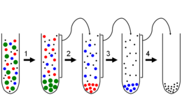
Differential centrifugation is a separation process in which cellular components are separated by several serial centrifugations at increasingly higher speeds. It takes advantage of the fact that centrifugation separates the cell components based on their size and density. Large and dense components migrate the fastest, which means that they settle out at relatively low speeds and form a pellet .
A cell homogenate is centrifuged (1.) at low speeds. The resulting pellet (green) consists of large, heavy components. The supernatant is removed and centrifuged at a slightly higher speed (2.). This process is repeated and the speed increased with each centrifugation. The following rules of thumb apply to the separation of cellular components: cell nuclei sediment during centrifugation at 1000 g for 5 - 10 min; Mitochondria at 10,000 g for 15 min and microsomes at 100,000 g for 1 h.
literature
- Alberts , Bray, Johnson, Lewis: Textbook of Molecular Cell Biology. 2nd, corrected edition. Wiley-VCH, Weinheim 2001, ISBN 3-527-30493-2 .
Individual evidence
- ↑ F. Lottspeich, H. Zorbas (Ed.): Bioanalytik. Spektrum Akademischer Verlag, Heidelberg 1998, ISBN 3-8274-0041-4 .
