White matter
| White matter | |
|---|---|
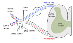
 The formation of the spinal nerve from the dorsal and ventral roots. (White matter labeled at right.) | |
| Details | |
| Identifiers | |
| Latin | substantia alba |
| MeSH | D066127 |
| TA98 | A14.1.00.009 A14.1.02.024 A14.1.02.201 A14.1.04.101 A14.1.05.102 A14.1.05.302 A14.1.06.201 |
| TA2 | 5366 |
| FMA | 83929 |
| Anatomical terminology | |
White matter is one of the three main solid components of the central nervous system. White matter tissue of the freshly cut brain appears white to the naked eye because of being composed largely of lipid. The other two components of the brain are gray matter and substantia nigra.
Structure
White matter is composed of bundles of myelinated nerve cell processes (or axons), which connect various gray matter areas (the locations of nerve cell bodies) of the brain to each other, and carry nerve impulses between neurons.
Cerebral- and spinal white matter do not contain dendrites, which can only be found in grey matter along with neural cell bodies, and shorter axons.[citation needed]
Function
The white matter is the tissue through which messages pass between different areas of gray matter within the nervous system. Using a computer network as an analogy, the gray matter can be thought of as the actual computers themselves, whereas the white matter represents the network cables connecting the computers together. The white matter is white because of the fatty substance (myelin) that surrounds the nerve fibers (axons). This myelin is found in almost all long nerve fibers, and acts as an electrical insulation. This is important because it allows the messages to pass quickly from place to place.
The brain in general (and especially a child's brain) can adapt to white-matter damage by finding alternative routes that bypass the damaged white-matter areas, and can therefore maintain good connections between the various areas of gray matter.
Unlike gray matter, which peaks in development in a person's twenties, the white matter continues to develop, and peaks in late middle age.[citation needed]
Location
White matter forms the bulk of the deep parts of the brain and the superficial parts of the spinal cord. Aggregates of grey matter such as the basal ganglia (caudate nucleus, putamen, globus pallidus, subthalamic nucleus, nucleus accumbens) and brain stem nuclei (red nucleus, substantia nigra, cranial nerve nuclei) are spread within the cerebral white matter.
The cerebellum is structured in a similar manner as the cerebrum, with a superficial mantle of cerebellar cortex, deep cerebellar white matter (called the "arbor vitae") and aggregates of grey matter surrounded by deep cerebellar white matter (dentate nucleus, globose nucleus, emboliform nucleus, and fastigial nucleus). The fluid-filled cerebral ventricles (lateral ventricles, third ventricle, cerebral aqueduct, fourth ventricle) are also located deep within the cerebral white matter.
Types of astrocytes
In 1983, M. C. Raff et al.[1] discovered that tissue samples originating from rat optic nerve contained two morphologically distinct types of astrocytes.
- So-called "Type 1 astrocytes" had a fibroblasts appearance and resided in both gray matter and white matter.
- "Type 2 astrocytes" has a neuron-like appearance and resided in white matter alone (Sherman, Chris).
Clinical relevance
Multiple Sclerosis (MS) is one of the most common disease which affects white matter. In MS lesions, the myelin shield around the axons has been destroyed by inflammation.
Changes in white matter known as amyloid plaques are associated with Alzheimer's disease and other neurodegenerative diseases. White matter injuries ("axonal shearing") may be reversible, while gray matter regeneration is less likely.
In November 2007, the Toronto based Centre for Addiction and Mental Health published a study in the Journal of Psychiatry Research showing that child molesters with pedophilia suffer from a "significant lack of white matter connecting six different areas of the brain all known to play a role in sexual arousal."[2][3]
The study of white matter has been advanced with the neuroimaging technique called diffusion tensor imaging where magnetic resonance imaging (MRI) brain scanners are used. As of 2007, more than 700 publications have been published.[4]
Notes
- ^ Raff MC, Abney ER, Cohen J, Lindsay R, Noble M (1983). "Two types of astrocytes in cultures of developing rat white matter: differences in morphology, surface gangliosides, and growth characteristics". J. Neurosci. 3 (6): 1289–1300. PMID 6343560.
{{cite journal}}: Unknown parameter|month=ignored (help)CS1 maint: multiple names: authors list (link) - ^ Brain wiring link to paedophilia
- ^ Pedophiles have less brain white matter: Toronto study
- ^ Assaf Y, Pasternak O (2008). "Diffusion tensor imaging (DTI)-based white matter mapping in brain research: a review". J. Mol. Neurosci. 34 (1): 51–61. doi:10.1007/s12031-007-0029-0. PMID 18157658.
