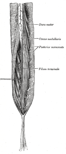
Conus medullaris
The conus medullaris (Latin for medullary cone ) is the tapering, cone-shaped caudal (rear or lower) end of the spinal cord . Its position in the spinal canal changes during the development of a person, as the spine grows longer than the spinal cord, and shows clear individual fluctuations. At the time of birth, the medullary conus is usually at the level of the third lumbar vertebra (L3), in adults it is approximately at the level of the first to second lumbar vertebra (L1 / L2).
As the ventriculus terminalis , also known as the V. ventricle , a dilatation containing liquor that is almost 1 cm long and 2-4 mm wide and connected to the central canal can be found within the lower end of the cone in the newborn . This of ependymal cells lined cavern provides a relic is that in humans after vacuolization, channeling and neuroepithelial lining of a first as caudal cell mass secondary applied tail region (in the amount of 31 somites ) remaining from incomplete regression in evolving spinal cord.
The pia mater immediately adjacent to the medullary cone continues caudally beyond its apex as the thread-like filum terminale (medullae spinalis) , which runs within the dural sheath and is connected to its lower end (approximately at the level of the 2nd sacral vertebra). This approximately 15 cm long filum terminale [internum] contains connective tissue and fat as well as glial cells, as well as (degenerated) neurons and nerve fibers. The dural sac is fused with the dorsal periosteum of the coccyx via its external continuation, the filum terminale externum [durale] , a short, coarse cord of connective tissue also known as the ligamentum coccygeum . In this way, the position of the spinal cord in its skins can be stabilized by tensioning the thread-like structure.
Beneath the conus medullaris, in addition to the filum terminale, the roots of spinal nerves run as cauda equina , the spinal column passage through which is deeper (see figure).
Blood supply
The conus medullaris is supplied with blood via three arterial spinal longitudinal vessels: the anterior spinal artery ( Arteriae spinalis anterior ) and the paired posterior spinal arteries ( Arteriae spinales posteriores ). Other, less prominent way of the blood supply are (Rami spinales) segmental flows over radicular branches spinal branches from the abdominal aorta , the fifth lumbar arteries ( Aa. Lumbar ), the lateral cross-arteries ( Aa. Sacrales lateral ), the iliac lumbar arteries ( Aa. Iliolumbales ) and the unpaired central cross artery Arteria sacralis mediana , the latter serving more to supply the cauda equina.
pathology
The cone syndrome is a collection of signs and symptoms that are associated with disorders or injuries in the area of the Conus medullaris.
In a spinal lipoma , infiltration of the conus or filum terminale can lead to a tethered cord .
Individual evidence
- ^ L. Coleman, R. Zimmerman, and L. Rorke: Ventriculus terminalis of the conus medullaris: MR findings in children . In: AJNR Am J Neuroradiol . Volume 16, No. 7, August 1995, pp. 1421-1426. PMID 7484626 . , as a pdf
- ↑ bra. Choi, RC. Kim, M. Suzuki, W. Choe: The ventriculus terminalis and filum terminale of the human spinal cord . In: Hum Pathol . Volume 23, No. 8, August 1992, pp. 916-920. doi : 10.1016 / 0046-8177 (92) 90405-R . PMID 1644436 .
- ↑ Benninghoff: Macroscopic and microscopic anatomy of humans, Vol. 3. Nervous system, skin and sensory organs . Urban and Schwarzenberg, Munich 1985, ISBN 3-541-00264-6 , pp. 105f and p. 174, respectively.
- ↑ Andreas Bickel, Roland Gerlach: Case book neurology . Thieme, Stuttgart 2009, ISBN 978-3-13-139322-7 , p. 57 .
