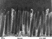
Microvilli
Microvilli (singular: microvillus, from the Latin villus ' villus ') are thread-like cell processes that increase the surface area of cells and improve the exchange of substances. Microvilli are mainly present in (animal) epithelial cells (e.g. in the intestine, kidneys, taste buds, uterus, egg cells) and form the brush border typical of these epithelia.
General properties
Microvilli are located on the (apical) side of the epithelial cell facing away from the basement membrane . These finger-shaped, brush-like protuberances have a reduced diffusion resistance for small molecules and are therefore extremely well suited for the absorption and release of substances in terms of their structure. These are transported along the stabilizing actin filaments , which run from the tip of the microvilli to their base. The transport is also supported by common rhythmic contractions or pumping processes.
structure
The finger-shaped microvilli are approx. 0.1 µm thick and, depending on the cell type, up to 2 µm long. Each microvillus contains a central bundle of actin filaments . They are held together in their shape by the proteins fimbrin and fascin . The actin bundle is connected to the lateral surface by myosin-I and to the cytoskeleton by spectrin . Each microvillus has an amorphous cap region at the apical end .
The microvilli are covered in a layer of proteins, glycoproteins, and sugar residues called glycocalyx . They determine antigenic properties. The glycocalyx also plays a role in cell recognition and adhesion mechanisms .
See also
literature
- Werner Müller-Esterl: Biochemistry - An introduction for physicians and natural scientists . 1st edition. Elsevier Verlag, Munich / Heidelberg 2004, ISBN 3-8274-0534-3 .
- RE McConnell, JN Higginbotham, et al. a .: The enterocyte microvillus is a vesicle-generating organelle. In: Journal of Cell Biology . Volume 185, Number 7, June 2009, pp. 1285-1298, ISSN 1540-8140 . doi : 10.1083 / jcb.200902147 . PMID 19564407 . PMC 2712962 (free full text).
