Cheyletiellose
| Classification according to ICD-10 | |
|---|---|
| B88.0 | Other acarinosis |
| ICD-10 online (WHO version 2019) | |
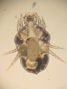
Cheyletiellosis is a parasitic disease of the skin that occurs mainly in rabbits and predators , but can also affect humans. It is caused by mites of the genus Cheyletiella .
Pathogen
The mites are up to 0.4 mm in size, have four pairs of legs and live on the host without leaving it through intermediate stages, so that the entire development cycle takes place on the affected animal. It takes about three weeks. The infection occurs through direct or indirect contact. Outside the host, female mites can survive for up to 10 days , whereas larvae and nymphs die quickly. Cheyletes have a rather broad host range. Even if the individual predatory mite species are preferred on some hosts, the host specificity is not very high. They can also temporarily infect humans.
| Art | Main host (s) |
|---|---|
| Cheyletiella blakei (Smiley, 1970) | Cats , foxes , humans |
| Cheyletiella parasitivorax (Mégnin, 1878) | Rabbits , cats, silver fox , dogs |
| Cheyletiella strandtmanni (Smiley, 1970) | Rabbits |
| Cheyletiella yasguri (Smiley, 1965) | Dogs, cats (rare), humans |
clinic
A Cheyletiellosis is highly contagious and plays a larger role, especially in larger holdings (breeding, pet shops).
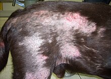
Since Cheyletes feed primarily on skin secretions and other harmless skin inhabitants, the infestation can also remain symptom-free. Typical small dry scales with or without itching can occur, especially in young animals . Miliary dermatitis can occur in cats . In the case of itching, severe skin changes can also result from self-harm. The disease can also affect humans and cause severe changes in the form of itchy papules on the abdomen or arms, which however heal after about three weeks without further contact with infected animals.
Even at low magnification, the white mites and the nits adhering to the hair are clearly visible. Since the mites are very mobile and live on the surface of the skin, a specimen with a transparent adhesive strip, which is then examined microscopically, is particularly suitable for detection .
To combat acaricidal substances, such as pyrethroids , amitraz , ivermectin , selamectin or doramectin , can be used.
literature
- Ch. Noli and F. Scarampella: Predatory mite . In: Practical Dermatology in Dogs and Cats . Schlütersche Verlagsanstalt, 2nd edition 2005, p. 236. ISBN 3-87706-713-1
Individual evidence
- ↑ Katrin Timm and Claudia S. Nett-Mettler: Pruritus in dogs (part 2) - Infectious and neoplastic causes. In: Kleintierpraxis , Volume 60, 2016, Issue 6, pp. 311–332.


