
Coat colors of dogs
The coat color of dogs , like the coat colors of other animal species, is controlled by different genes . A coat color that looks the same can result from very different combinations of genes that cannot be recognized by the phenotype ( polygeny ).
Control of dye production
There are two colorants (melanins) found in fur : black eumelanin and red pheomelanin . All coat colors known from dogs are caused by different distribution of these two pigments in the coat. Some color genes control when and where these dyes should appear in the fur and skin. Of these control loci, the extension locus (E) and the agoutilocus (A) have been researched best .
If a gene affects the control of melanin synthesis, i.e. determines whether and where which melanin should be produced, this can often be recognized by the fact that all colorants can be produced, but appear in different places.
Note: In the articles on the gene loci there are sections with their own headings on the dog.
| Cross-species gene locus | chromosome | Mutation, allele | Colours |
|---|---|---|---|
| Agouti | CFA24 | fawn / sable A y | Deer -colored, sand-colored, yellow or reddish with darker markings e.g. B. on the nose; black hair partly scattered |
| Agouti | CFA24 | wolf sable a w | Wild-colored animal in which the individual hair is banded |
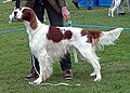
| Agouti | CFA24 | a t | Black with red branding or tan markings or tan with black or gray saddle |
| Agouti | CFA24 | a | Recessive black |
| Extension | CFA5 | E M | Black mask |
| Extension | CFA5 | E. | Black fur areas possible |
| Extension | CFA5 | e | Fawn-colored , sandy-colored, yellow or reddish all over the body . Only pheomelanin can be produced in the fur; Skin black if not lightened by a gene for leucism or albinism |
| K locus (see melanism in dogs) (beta-defensin 103 (CBD103) gene) | CFA16 | k B | Dominant black color |
| K-Locus (see currents in dogs) | CFA16 | k br | Areas that would get their color from pheomelanin at k y appear brindle |
| K locus | CFA16 | k y | Phaeomelanin can be produced |
| In addition to the data on the genes, the table also shows what the colors are called when the gene affects the three basic colors of the genes. The genes of a locus are indicated in their order of dominance , the most dominant first. |
|||
The effect of the three gene loci on one another
In the K locus, k y is the most common allele and at the same time the wild type. When this allele is present, the alleles of the agouti and extension locus result in the following colors.
| Color variations at k y | fawn / sable a y | wolf sable a w | a t | a |
|---|---|---|---|---|
| E M E M , E M E or E M e | Light brown with a black mask on the face | Wild colors with a black mask on the face | Black with red burn, mask stays black | black |
| EE or Ee | Light brown without a mask | Wild colors without a black mask on the face | Black with red burn | black |
| ee | Light brown without a mask | Light brown without a mask | Light brown without a mask | Light brown without a mask |


The allele for flame k br of the K locus is dominant over the allele k y . So the flamed drawing is expressed when the allele combinations k br k br or k br k y are present. Depending on the genes on the agouti and extension locus, different markings can occur. The fur areas that are light brown at k y are each flamed light brown black, while the black fur areas remain unchanged black. Here, too, the allele e of the extension locus is epistatic via the flame k br and the dogs with the gene combination ee are light brown regardless of the K locus.
The allele k B for dominant black staining is the dominant allele of the K locus. The allele combinations k B k B , k B k br or k B k y all lead to the same appearance. Of the other gene loci, only the allele e of the extension locus is epistatic over the dominant black color. If k B is present at least once, a dog is black with almost all gene combinations, only ee at the extension locus leads to a light brown coat color, whereby the nose can still be black due to an allele k B. Dogs with the dominant allele E, in which the recessive allele b is homozygous on the B locus (TYRP1 gene), have a brown nose and mostly brown fur.
| Color variations at k B k B , k B k br or k B k y | a y , a w , a t and a in any combination |
|---|---|
| E M E M , E M E, E M e, EE or Ee | black |
| ee | Light brown without a dark mask |
Albinism spectrum: mutated enzymes of melanin synthesis
To produce the two melanins, a number of different enzymes , structural proteins and transport mechanisms must work together properly in the dye-producing cell, the melanocyte . Mutations in the genes of the substances required for this mean that the animals concerned are not able to produce melanin or that they can only produce a small amount of melanin. Even lightening of the coat color is often due to changes in the enzymes involved in melanin synthesis. Mutations at the beginning of the melanin synthesis pathway affect both the red and the black dye. If the black and the red dye are lightened to different degrees, this is often because the gene intervenes towards the end of melanin synthesis, where the synthesis pathways of eumelanin (black) and pheomelanin (yellow, brown) have already separated.
Some mutations in this area, such as the merle gene , cause toxic intermediate products of cell metabolism to accumulate in the melanocytes, causing the cells to die.
| Cross-species gene locus | chromosome | Mutation, allele combination | Designation, description of the appearance |
|---|---|---|---|
| Albino locus (C), ( oculocutaneous albinism type 1 ), tyrosinase gene | CFA21 | ? | No mutations are known (2007) that affect the color of the animals concerned. The white color in Doberman Pinschers, Lhasa Apso and Pug are not due to mutations in the tyrosinase gene. |
| Braun locus (tyrosinase related protein 1 (TYRP1), oculocutaneous albinism type 3 ) | CFA11 | B. | Eumelanin is black (black dogs, dogs with black markings, black skin in monochrome light brown dogs) |
| Brown locus ( oculocutaneous albinism type 3 ) | CFA11 | Braun gene b (3 variants: b s , b d or b c ) | Eumelanin: Black is lightened to brown, the skin on the nose, around the eyes and under the feet is also lightened to brown. Phaeomelanin is not lightened. The fur of dogs with the genotype e / e remains unchanged, but the skin color on the nose, eyelids and pads is lightened from black to brown. |
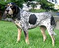
| Silver locus | CFA10 | Merle factor , pure breeding (MM) | Merles, tigers, very light-colored animals, mostly eye damage or deafness |
| Silver locus | CFA10 | Merle factor , mixed mix (Mm) | Merle, Tiger, healthy; mainly eumelanin is lightened. |
| Silver locus | CFA10 | no merle factor (mm) | Not lightened, healthy |
| Melanophilin gene ( MLPH ) | CFA25 | D. | Not lightened |
| Melanophilin gene ( MLPH ) | CFA25 | Dilute gene (d) | (Engl. Whitens black to "Blue" blue ), which one is actually gray. Phaeomelanin is hardly influenced by the gene. |
| Melanophilin gene ( MLPH ) | CFA25 | Dilute gene (d 2 ) | Brightens black (Engl. To "Blue" blue ), which is actually a gray. Phaeomelanin is hardly influenced by the gene. Skin problems |
| unknown A | unknown C | P | Not lightened |
| unknown A | unknown C | p | Eumelanin and Phaeomelanin are brightened to white |
| unknown A | unknown C | G | Gradual graying (like mold) |
| unknown A | unknown C | G | Not lightened |
| unknown A | unknown C | C. | Not lightened |
| unknown A | unknown C | c ch | Eumelanin and Phaeomelanin are lightened |
| unknown A | unknown C | c a | Complete albinism |
| unknown A | unknown C | I. | Not lightened |
| unknown A | unknown C | mixed: Ii | Eumelanin is not lightened, Phaeomelanin is lightened |
| unknown A | unknown C | i | Eumelanin is not brightened, pheomelanin is absent |
Examples of the effects of genes on the albinism spectrum
| Starting color | Lightened by the brown gene (bb) | Lightened by Merle Factor (Mm) | Lightened by the dilute gene (dd, dd 2 or d 2 d 2 ) | Lightened by ii |
|---|---|---|---|---|
| black | brown | Blue merle | Blue (looks gray) | Unchanged: black |
| Black with red burn | Brown with fire | Blue merle with red fire | Blue (looks gray) with red burn | Black with white burn |
| Light brown (due to pheomelanin) with a black snout | Light brown with a brown muzzle | Unchanged: light brown with a black snout | Unchanged: light brown with a black snout | White |
| brown | Brown, genetically identical to the original color | Red Merle (wrongly shown with fire) | Typical color of the Weimaraner (Isabell) | Unchanged: brown |
Leucistic color genes
In leucism, the dye-forming cells ( melanocytes ) do not migrate out of the neural crest during embryonic development , in fewer numbers than usual or too late. The following gene loci were known to be the cause of leucism: endothelin receptor B gene (EDNRB), the paired box gene 3 (PAX3), SOX10, the microphthalmia-associated transcription factor (MITF), c-Kit and the steel locus (coded MGF). With complete leucism, the affected animal is completely white and can have normal colored, slightly brightened, blue or red eyes. Less pronounced leucism leads to piebald animals, to white markings on the head and legs or to animals with white prickly hair in otherwise normal-colored fur.
Every check pattern is possible on every basic color.
Similarly, there are at piebald considerable individual differences in the expression of spotting: Most rich at the same Scheckungsgen the variants of entirely white dogs to dogs, although they carry the Scheckungsgen but not outwardly appear pied or have only a discreet little patch because this gene .
White markings on the face and legs are also due to leucism in most species.
| Cross-species gene locus | chromosome | Name of the mutation | General name |
|---|---|---|---|
| MITF | CFA20 | S. | No check |
| MITF | CFA20 | s i | Irish spotting |
| MITF | CFA20 | s p | Piebald |
| MITF | CFA20 | s w | White with colored head (extreme white) |
| unknown A (leucism) | unknown C | T (ticking) | White with brown panels and brown speckles, brown mold with panels, black mold with panels (belton, ticked or, depending on the basic color, bluetick, redtick, red or blue roan) |
| unknown A (leucism) | unknown C | t | Without white |
| unknown A (leucism) | unknown C | R (roan) | A roan gene was postulated for a dog with white hair in its colored fur. Presumably, however, it is identical to the gene T (ticking) |
| unknown A (leucism) | CFA9 | HH | Fatal in early embryonic development |
| unknown A (leucism) | CFA9 | Hh (Harlequin) | Harlequin spotting if the merle gene is also present at least once |
| unknown A (leucism) | CFA9 | hh (no Harlequin) | No Harlequin check |
| unknown A (leucism) | unknown C | not named | White underside in both homozygous and heterozygous forms of the gene |
s p Piebald
b w white with colored head
T white with black panels and black speckles

T - mixed light and dark hair

Boxer: s i s i ( wrong color )

Boxer: Ss i

Great Dane: H (Harlequin gene) and Mm (Merle gene heterozygous)
Border Collie: Although they have the white in the same places as heterozygous boxers with the gene combination Ss i , the color is caused by a different gene.

Dalmatians: It is believed that the Dalmatian spots are caused by two genes, one T (ticking) and an unknown additional gene.

At birth, Dalmatians are still white. The spots only appear after a few days.


.jpg)

Relationship between coat color and health and behavior
Coat color can also affect a dog's health and behavior. This is because the genes that cause different coat colors often play a role in other processes in the body ( pleiotropy ).
health

Visual Impairments: In humans and all animal species, complete albinism results in red eyes and a visual impairment due to a lack of melanin in the eye. Weakened forms of albinism, such as those caused by the dilute gene , can, depending on the degree of severity, lead to minor visual impairments or have no noticeable effects on vision. In the case of Dilutegen, the dog is not known to have any visual impairment. Leucism can cause visual impairment similar to albinism. In the case of Merle syndrome , smaller eyeballs and malformations of the lens also occur if the dog has the merle gene twice ( homozygous ), since two parents, both of which have the merle gene, were mated. Of the heterozygous Merle dogs, 2.7% are one-sided, 0.9% are completely deaf (see Merle factor ).
Deafness: Leucism is often associated with deafness. 12–22% of Dalmatians are deaf in one ear and 5–9% in both ears. There is a connection with the color genes, because animals with blue eyes are significantly more likely to be deaf, animals with plate markings are less often deaf. The Merle factor belongs more to the albinism spectrum, but genetic carriers of the gene are in many cases deaf in one or both ears.
Skin Diseases: The dog's dilute gene (MLPH) is often linked to hair loss and changes in the hair roots .
behavior
For the cocker spaniel it has been shown in several studies that animals colored in gold or reddish brown are the most aggressive, black cocker spaniels are in the middle and dogs of the blue or red gray colors are the least aggressive.
Nomenclature in the FCI
In the FCI , the largest international organization for dog breeding, a nomenclature has existed since 2009 for the designation of the hair colors of dogs. This was published by Bernard Denis in the book Coat Colors in Dogs . The descriptive part was summarized and approved as the Standardized Nomenclature of Coat Colors in Dogs by the FCI Scientific Commission in July 2009 as a reference. It should be used for revisions and new versions of breed standards .
Individual evidence
- ↑ Genomia: Locus A
- ↑ Genomia: coat color of dogs http://www.genomia.cz/de/dogcolor/
- ↑ Sheila Schmutz 2016: Dog Coat Color Genetices: http://homepage.usask.ca/~schmutz/dogcolors.html
- ↑ Genomia: Locus B: nose color http://www.genomia.cz/de/test/locus-b-dog/
- ↑ Genetics of coat colors in dogs - modifier genes
- ↑ Anima Labs: S-locus
- ↑ a b c L.A. Clark, AN Starr, KL Tsai, KE Murphy: Genome-wide linkage scan localizes the harlequin locus in the Great Dane to chromosome 9. In: Gene. 418 (1-2), 2008 Jul 15, pp. 49-52. Epub 2008 Apr 16. PMID 18513894
- ↑ Simone G. Rak, O. Distl, I. Nolte, J. Bullerdiek: Molecular genetic investigation of the congenital deafness in the Dalmatian. In: Society for the Promotion of Cynological Research (ed.): Circular 13 (2001) pp. 41–46.
- ↑ Joaquín Pérez-Guisado, Andrés Muñoz-Serrano, Rocío López-Rodríguez: Evaluation of the Campbell test and the influence of age, sex, breed, and coat color on puppy behavioral responses. In: The Canadian Journal of Veterinary Research. 72, 2008, pp. 269-277.
- ↑ Standardized Nomenclature of coat colors in dog
Article sources
Unless otherwise stated, information in this article has been obtained from the following two sources:
- SM Schmutz, TG Berryere: Genes affecting coat color and pattern in domestic dogs: a review. In: Anim Genet. 38 (6), 2007 Dec, pp. 539-549. PMID 18052939 [1]
- Sheila Schmutz: Dog Coat Color Genetics. Status 7/2008.
Web links
- Fur colors using the example of Eurasier
- Article series on coat colors by Anna Laukner, published in the Swiss dog magazine (overview of all 12 articles with links)