
Check
There is a piebald in humans and most animal species. It is a weakened form of leucism in which the body is not all white, only some parts of the body have white spots.
Leucist check
Formation of the spots
There are several genes whose mutation can lead to leucism and spotting. These include the endothelin receptor B gene (EDNRB), the paired box gene 3 (PAX3), SOX10, the microphthalmia-associated transcription factor (MITF), c-kit and the steel locus (encoding MGF).
In an early embryonic precursor cells of the wandering Schwann cells , the ganglion cells of the auditory nerve and the precursor of melanocyte from above the neural tube lying neural crest from. The spotting arises from the fact that one of the genes that influence the emigration of melanocytes is mutated and therefore not as many cells emigrate and they do not reach all parts of the body. Different spotting patterns can arise depending on the type of mutated gene. Such a mutation either only influences the melanocytes or also influences the other types of progenitor cells. Spotting and leucism are usually inherited dominantly , but depending on the species and subspecies, they can also be inherited as an intermediate , co-dominant or recessive trait.
The precursor cells of melanocytes are called melanoblasts . If they fail completely, the affected animal turns white because the cells that produce the melanin are missing . This is called leucism. If fewer cells have emigrated and are evenly distributed in the body, the animal becomes a bit lighter than wild-colored animals, but looks similar (lightened). In piebald animals, the melanocytes do not get everywhere, while in other places there are normally many melanocytes.
Among the Leucistic color genes include the piebald gene in humans the piebaldism caused.
Eye color and visual impairment
The complete absence of melanin in the iris of the eye leads to a red eye color, an almost complete absence leads to blue eyes. When the amount of melanin is reduced just a little, the eyes are light brown. Depending on the location of the spots and the type of underlying leucism, piebald animals can have blue to normal-colored eyes, sometimes they also have two different eye colors ( iris heterochromia ). The melanocytes also affect the development of the optic nerves , so leucistic animals often have a visual impairment equivalent to the visual impairment of animals with albinism .
Hearing loss and deafness
If the spotting is caused by a gene or a certain variant of an allele that influences the precursor cells of the ganglion cells of the auditory nerve , whereby there may also be a lack of melanocytes in the stria vascularis , the affected animal becomes hard of hearing or deaf. The melanocytes do not form pigments in the stria vascularis, but connect two other layers of tissue that can only fulfill their function in this way. After initially undisturbed development of the inner ear , degenerative changes occur in the cochlea in the first days of life . The severity of changes in the inner ear can vary.
Nervous system disorders
The Schwann cells envelop the axons of the nerve cells . They enable a much faster conduction of excitation in the neurons of the deuterostomia , which also include mammals and birds , than in the nerve cells of the protostomia , which include insects . If the mutated gene affects the migration of these cells, the nervous system cannot work properly.
Severe deformities
Individual Scheckloki also lead to further severe malformations of the affected animals in the embryonic development, which can go so far that animals that have the mutated gene on both homologous chromosomes , i.e. are homozygous , die in the womb or shortly after birth.
No deformities
However, there are also harmless variants of most check loci that only lead to white spots.
Leucistic spotting in various animal species
human
People can also be piebald, which means that they have light spots on their skin. The spots are usually sharply defined. This is called piebaldism . In 90% of the cases, those affected have a light-colored forelock. The underlying mutations affect the gene coding for the cKIT receptor on chromosome 4 in 75% of cases. The transmembrane cKIT receptor, with its ligand, the growth factor cKIT, is involved in the regulation of melanocyte proliferation and migration.
While most people with piebaldism are healthy, Waardenburg syndrome is a form of spotting in humans that is associated with hearing loss and eye defects.
Vitiligo and tuberous cerebral sclerosis can also lead to white spots on the skin, but are not spotted in the sense of the word treated here.
Pigs
In isolated cases, wild boars with white spots were known. These animals are descendants of hybrids , so they can be traced back to runaway piebald domestic pigs that were mated by wild boars . Under today's conditions, they can hardly hold their own in the wild because human hunters can see spotted animals more easily. The spotting gene of wild boars is inherited recessively , so that the conductors are not under selection pressure and can pass it on. Both the pink color of today's domestic pigs and the piebalds and the belt markings of some breeds can be traced back to individual mutations of the kit locus.

Black and white belt drawing on domestic pigs

Spotting with small black spots

Hunter with a shot young wild boar with belt drawing


Domestic cattle


Pied domestic cattle breeds roughly sorted according to the type of piebald:
Largely white head, white belly, white stripes on the back line: Abondance (cattle) , Ennstaler Bergschecken , Simmental cattle , Hinterwälder cattle , Montbéliard (cattle) , Pustertal piebalds , Hereford cattle
Only the belly and back are white: Evolèner
Speckled: Ayrshire cattle , Holstein cattle , Maine Anjou , Red Holstein lowland cattle , Black & white lowland cattle , Vorderwälder cattle
Speckled: Gir (zebu) , Norman (cattle)
Domestic goats

Examples of spotted breeds are: Colorful Dutch goat , Boer goat , Tauernscheckenziege , Thuringian forest goat , Walliser black-necked goat , West African dwarf goat
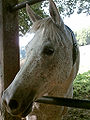
Horses
In spotted horses , different genes can be responsible for the color .
The American Paint Horse Association, which deals with the targeted breeding of special colors, distinguishes the following colors:
.JPG)
Tobiano : The legs are usually white or have white markings, the head usually does not have more white markings than unchecked horses. The eyes are mostly dark. The white usually crosses the topline somewhere and the spots lead from top to bottom.
Frame Overo : The back line is always colored throughout. The face usually has larger white markings while the legs are mostly dark, but can sometimes have white socks. The color varies between almost white and almost colored types. Homozygous animals are completely white and not viable.
Splashed White Overo : The horse looks like it has been dipped in white paint. The back and ears are colored, the legs and lower half of the body are white. Many Splashed White Overos are deaf.
Sabino Overo : The horse usually has four white legs, the white then spreads upwards in small speckles. Some Sabinos have blue eyes, others have brown eyes. If the pattern is only slightly pronounced, they have only white legs and a blaze. Others are almost completely white or very easily confused with the other forms of spotting. The Sabino Overo spotting is probably due to several different genes. Sometimes the "lethal white" gene also appears in Sabinos
Tovero: Mixture between Tobiano and Overo, sometimes Sabinos too.
dog


There are many different piebald and mottled genes in dogs .
Almost 10% of Dalmatians are deaf in one or both ears. Since the deafness is caused by a piebald gene, you cannot breed away this risk of deafness without renouncing the typical Dalmatian color.
Red fox
It is very rare to find red foxes with a piebald with large, white areas of follicle and skin. It was observed in connection with a general brightening (Weißling) and in the breeding breed platinum fox . In the fox populations in Ireland about 5% of the animals have spots in the lower wool hair (+ skin) with small, widely scattered white spots. The stain is covered by the long top hair. It is not known how these spots behave in inheritance compared to the normal red fox fur color.
Domestic cat
The spotting of the house cat is caused by an autosomal gene S, which can be assigned to the c-Kit locus and is subject to an incompletely dominant inheritance. Animals with this gene can look very different. There are smooth transitions from colored animals with small white-haired spots in the chest area, or along the middle of the belly to completely white cats. In cats with the mutated gene, the lack of melanoblasts sometimes causes hearing loss or deafness.
Birds


In birds, the leucistic spotting is caused in the same way as in other animals. The complete pigmentation in the head area at the Elster can be taken as an indication that there is no impairment of the sensory organs. On the other hand, the shown purple Grackel , which differs greatly from its typical appearance , also shows a pronounced leucistic spotting in the head area. Poor eyesight due to a lack of melanin in the eyes is much more dangerous for the life of flying animals than for animals living on the ground. Flying is one of the skills that makes the greatest demands on the sensory performance of the eyes and the optical evaluation of the sensory stimuli. As a result, birds with visual impairments are often injured because of failed landing maneuvers and overlooked mesh and barbed wires.
Other spots
Spots attributed to albinism
The coloring of the point cats and the Russian rabbits is due to albinism (OCA1). A heat sensitive tyrosinase is present.
In the pink eye series (P) there is an allele p m whose color forms a mosaic between wild type and lightening.
No alleles are known on the brown locus (OCA3) in pigs with the exception of the B k allele , which leads to the development of brown spots on a red background.
Spots from the silver gene
The merle factor in domestic dogs is a mutation of the silver locus , which, in addition to irregular white spots in the fur, can lead to malformations of the eyes such as the lack of a lens or reduced eyeballs. In addition, malformations of the inner ear occur with deafness or hearing loss.
Wind-colored horses often have white apples in their fur, which is caused by a mutation in the same genetic locus.

Blue merle Catahoula Leopard Dog

Black horse with silver gene and apple drawing
Head of a gray fly


Apple mold and fly mold
Horses with the gray gene often have apple markings (apple mold) or small black dots in their fur (fly mold.)
Tiger piebald and varnish roan
Tigerscheckung : Schabrackeschecke, Tigererschecke, Varnisch Roan.
The gene for the tiger piebald complex is a mutation of the TRPM1 gene ( Transient Receptor Potential Cation Channel, Subfamily M, Member 1), which causes the expression of the gene in homozygous animals to be reduced to 0.05% of the level in normal-colored horses is. The expression rate of heterozygous animals lies in between. It is assumed that the reduced expression of the gene disrupts both the function of the bipolar cells and that of the melanocytes, so that both the night blindness of the homozygous Appaloosas and the typical Appaloosa color are thereby caused.

Volltiger

Saddleback tiger with small saddle pad (Appaloosa)

Varnish Roan


Fur drawings
In most animals, the agouti locus and the extension locus play a role in the development of typical spot markings. The stripes of the striped cats depend on the tabby locus, the K locus is responsible for the flow of the dogs.
Articles about patterns:
Spotting through different masking of genes

With some genes, an allele of the respective gene is switched off by imprinting or X-inactivation . This can lead to piebalds that have similar distributions to black and white piebald animals, but alternate between two colored colors.
Mouse: Viable Yellow
In mice which have the gene "Viable Yellow" on one allele of the agouti locus and the gene for black color on the other allele, individuals with black and yellow speckles can occur due to different imprinting.
Cat: Tricolor cats and tortoiseshell cats
The tortoiseshell pattern consists of red and black areas of fur and occurs almost exclusively in female cats. The gene for red color is on the X chromosome and is randomly switched on or off in heterozygous cats in different areas of the coat ( X inactivation ). This creates a normal-colored red check.
See also
- Leucism
- Albinism
- Coat colors of horses
- Genetics of horse colors
- Domestic rabbit genetics
- Coat colors of the cat
- Coat color
- Variegation in plants
swell
- E. von Lehmann: Some remarks on the spotting of the red fox (Vulpes vulpes crucigera Bechstein, 1789). In: Journal for Hunting Science. Volume 36, Number 3, September 1990, pp. 195-197.
- Petra Keller: Studies on the development of the early acoustic evoked potentials (FAEP) in cats for use in basic research and clinical application. Inaugural dissertation . University of Veterinary Medicine Hannover, 1997
- Richard A Spritz, Stuart A Holmes, Peter Itin, Wolfgang Küster: Novel Mutations of the KIT (Mast / Stem Cell Growth Factor Receptor) Proto-Oncogenes in Human Piebaldism. In: Journal of Investigative Dermatology . (1993) 101, pp. 22-25.
- TT Patterns, TO Patterns: The Genetic Equation of Paint Horses.
- J. Schäfer: Piebaldism. In: J. Gille, M. Wolter, R. Kaufmann; Nude dermatol. Frankfurt dermatologist evening on November 2, 2005. Stuttgart 2005.
- D. Dausch, W. Wegner, W. Michaelis, I. Reetz: Eye changes in the dog's Merle syndrome. In: Graefe's Archive for Clinical and Experimental Ophthalmology. Volume 206, Number 2, June 1978.
- Rainer Brinks: Defective Merle Factor. 2001.
- Anja Wriedt: Studies on the inheritance of colors in Japanese gulls (Lonchura striata f. Dom.) With special consideration of the manifestation of cataracts in the leucistic color variant. Dissertation . Hanover 2001.
Individual evidence
- ^ S locus (Piebald, white spotting). In: laboklin.de. Retrieved December 30, 2016 .
- ↑ a b c Saskia Kristina Hogreve: Studies on the hearing ability of New World camelids with special consideration of the iris pigmentation. Dissertation, Justus Liebig University Giessen, 2003.
- ↑ Sheila Schmutz 2014: Spots and White Markings