Albinism
| Classification according to ICD-10 | |
|---|---|
| E70.3 | Albinism |
| ICD-10 online (WHO version 2019) | |

Albinism (from Latin albus 'white') is a collective term for congenital disorders in the biosynthesis of melanins (these are pigments , i.e. dyes) that affect the resulting lighter skin , hair or coat color and eye color , but also on affect other characteristics ( polyphenia ). Affected animals are called albinos , affected people usually prefer the more neutral term "people with albinism". People with albinism get sunburn more easily and therefore more easily skin cancer . In addition, with complete albinism, visual acuity and spatial vision are impaired. The term Noach syndrome is also found here and there .
Albinism usually follows a recessive inheritance and occurs in humans worldwide with an average frequency ( prevalence ) of 1: 20,000. Clusters can be found above all in Africa with a prevalence of 1: 10,000 and higher. The light skin color of Asians and Europeans is due to albinism type OCA 4 , the blond hair and blue eyes of Europeans to OCA 2 and another gene.
In mammals, including humans, albinism occurs with lightened eyes, skin, and hair or coat color for the same reasons, since their dye synthesis is very similar. In other animal groups there are other dyes in addition to melanins and the use of the term albinism is inconsistent there. In birds, blue and green colors and shimmering play of colors are created by feather structures in conjunction with melanin. Yellow, orange and red colors are mostly due to carotenoids and pteridines . In reptiles, amphibians and fish, green and blue colors, a silvery shimmer or metallic sheen are created by purines that reflect light. All of these dyes can fail due to mutations.
Albinism in humans
Appearance and Symptoms

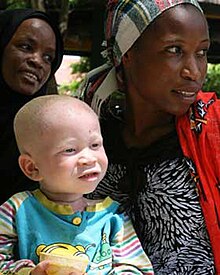
Even people whose bodies cannot produce any melanin at all, i.e. who are completely albinotic, are not extremely noticeable in Central and Northern Europe, as skin, hair and eyes that are lightened due to partial albinism are the norm as an adaptation to the lower solar radiation. Complete albinism results in pink skin, white-blonde hair, and pinkish-blue eyes in humans . People with less pronounced albinism are not always clearly recognizable as such by their appearance. They may look lighter than family members without albinism, but mostly a residual function of melanin production is still preserved, so that there are also dark-skinned people with albinism who have clearly brown skin and light brown eyes.
While most people with albinism have lighter eyes and hair colors than their non-albinotic blood relatives (oculocutaneous albinism, OCA), there are also cases of albinism in which the symptoms are limited to the damage to the eyes while they look normal on the outside (ocular Albinism, OA).
Skin color
People with albinism have lightened skin. They get easily sunburned and thus have a higher risk of skin cancer . The skin color of whites is lightened as an adaptation to the lower solar radiation outside the tropics, among other things by mutations in the albinism genes, but not to the extent that this results in recognizable eye damage.
Symptoms related to vision
In purely ocular albinism and in all forms of complete or almost complete oculocutaneous albinism, there is a pronounced complex of symptoms in the eyes . The color perception is normal, however, since albinism has no influence on the formation of rhodopsin .
Lightening of the eye color
The human eye colors of the iris can vary from dark brown to light brown and green to gray and blue or, if the iris is pigmentless, appear red because the blood-red vessels shine through.
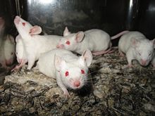
Albinism causes lighter eye color. Complete albinism leads to light blue, almost pink eyes as shown in the picture above, regardless of which eye color the person concerned would have without their albinism, which is very rare in humans. When very little melanin is produced, the eyes are blue. If there is less pronounced albinism, in which the body can still produce noticeable amounts of melanin, the eyes are correspondingly less brightly colored.
Photosensitivity
If the body can hardly or almost not produce melanin and it is therefore not present in the eye, or only to a very small extent, the iris becomes transparent to a certain extent and can be transilluminated with appropriate light. In less pronounced cases, the pigment defects are more likely to be found in the area of the iris root. This transparency is also evident when shining into the eye through red light reflections. Typical for people with severe albinism is therefore a pronounced sensitivity to glare ( photophobia ), which is why they usually wear sunglasses outdoors .

Disturbances in spatial vision
Melanin also plays a role in the development of the optic nerves . Normally, the field of vision in humans is evenly divided between both halves of the brain - each hemisphere has its side and both eyes deliver the part of the image that belongs to this side ( visual pathway ). By comparing the two images, each half of the brain can calculate the distance between the objects and assign it spatially ( stereoscopic vision ). In people with albinism, a larger proportion of the optic nerves crosses to the opposite side of the brain, which results in a loss of the physiological proximity of homologous retinal areas and images that belong together are not always processed on the same side.
In addition, there is usually ocular nystagmus (eye tremors) of varying degrees of severity, often accompanied by manifest strabismus (strabismus). In a series of tests of 37 patients, all had nystagmus of varying degrees, only four of them did not squint. The circumstances mentioned therefore often result in a lack of or at least significantly restricted spatial vision .
Decreased visual acuity
The fovea ( fovea ), the retina of sharpest vision is anatomically not fully formed when albinism because their development is also influenced by melanin. It is either not developed ( aplasia ) or develops only incompletely ( hypoplasia ).
Due to the nystagmus and the organic situation, the visual acuity (visual acuity) in a fully developed albinism is seldom more than 0.1, rather less, and can reach about 0.5 if the anomaly is less severe. The visual impairment varies greatly within the same type. Because of the existing pigment defects in the iris, contrasts between light and dark areas in the room can often only be seen indistinctly.
In addition, people with albinism are often unable to focus the eye at different distances ( accommodation ), and many people are nearsighted or farsighted .
Symptoms related to hearing
Albinism deafness syndrome is very rare .
treatment
Albinism does not affect people's mental development. Therefore, despite the non-treatable metabolic defect , they can lead a largely normal life with the help of (magnifying) visual aids , tinted glasses or contact lenses and appropriate skin protection.

Physiology of Albinism
Melanin synthesis
The dye melanin is produced by dye- producing cells, the melanocytes . The precursors of the embryo's melanocytes , the melanoblasts , migrate from the neural crest to the epidermis of the skin, hair follicles, and various other organs during pregnancy in the early fetal period . Once in the skin, the melanoblasts differentiate into melanocytes and form numerous cell processes through which they pass the melanin on to the keratinocytes . The number of melanocytes in blacks is the same as in whites, and someone with albinism also has a normal number of melanocytes. The color of the skin is determined by the amount and quality of the pigment melanin formed, not by the number of these cells.
Melanocytes contain melanosomes, small membrane-enclosed vesicles in which the pigment melanin is produced. In terms of their function, they are very similar to the lysosomes (cell organelles that are used for digestion within the cell), because both contain substances that are dangerous for the cell and therefore must not come into contact with the rest of the cell. The lysosomes contain protein-dissolving enzymes ( proteases ) and the melanosomes contain intermediate products of melanin synthesis such as quinones and phenols , which can damage the cell membranes.
In order to produce melanin, various enzymes are needed which, one after the other, help to build melanin ( gene chain ). When one of the enzymes in this metabolic pathway stops working, albinism occurs. The formation of eumelanin in the melanosomes begins with hydroxylation of the amino acid (AA) L-tyrosine by the membrane enzyme tyrosinase . In addition to this key enzyme, two other membrane-based enzymes DHICA oxidase and Dct are required so that eumelanin can be formed.
Molecular genetic classification of albinism
Although differences in the appearance of people with albinism were described early on, it was believed that albinism was due to changes in a single gene. It was not until the family described by Trevor-Roper in 1952, in which both parents were affected by albinism and still had normally pigmented children, that there was an initial indication of the genetic heterogeneity of this disease. Both parents were homozygous in this case for gene mutations that led to albinism. However, these concerned different genes, so that the children were heterozygous (mixed-breed) for each of the two mutations and were therefore not clinically affected by albinism.
First, albinism was classified according to external appearance . Later it was possible to prove whether tyrosinase - an enzyme necessary for melanin production - was present. With the possibility of identifying some genes responsible for the OCA, a molecular genetic classification was finally established. It was found that the different phenotypes are not always due to mutations in different genes, but often represent different manifestations of various mutations in one gene. Clinical differentiation remains difficult, as the mutation ( genotype ) causing the disease cannot be inferred from the appearance (phenotype) .
Four types of oculocutaneous albinism are known. Oculo-cutan is made up of (Latin) oculus: eye and cutaneus: relating to the skin. In ocular albinism, only the eyes are visibly changed.
| Albinism type | gene | Gene product | function | chromosome | Appearance | health |
|---|---|---|---|---|---|---|
| Oculocutaneous albinism type 1 , abbreviated OCA 1 | TYR | Tyrosinase | Enzyme in the synthesis of melanin | Chromosome 11 (11q14-21) |
OCA1A: Complete albinism, pink skin that does not tan, light blue eyes with a pink sheen OCA1B: incomplete albinism |
OCA1A: visual impairment with a visual acuity below 10% OCA1B: Visual impairment variable, visual acuity below 10% or not recognizable |
| Oculocutaneous albinism type 2 , or OCA 2 for short | OCA2 | P protein, a membrane protein of the endoplasmic reticulum | Transport of tyrosinase out of the endoplasmic reticulum | Chromosome 15 (15q11-13) | Responsible for blonde hair and blue eyes in Europeans
OCA2: variable: complete albinism up to barely noticeable lightening, sometimes with dark nevi (moles, birthmarks) on the light skin |
Europeans: healthy, no noticeable visual impairment
OCA2: Visual impairment variable Sometimes associated with Prader-Willi syndrome (PWS) or Angelman syndrome , as the genetic loci of both hereditary diseases lie next to one another. |
| Oculocutaneous albinism type 3 , or OCA 3 for short | TYRP1 | ( TYRP1 ) | DHICA oxidase, DHICA polymerase | Chromosome 9 (9p23) | In black people: brown or red-brown skin, brown hair, green-brown to brown eyes | no recognizable visual impairment |
| Oculocutaneous albinism type 4 , abbreviated OCA 4 | SLC45A2 | MATP | Transporter for the proteins of the melanocytes | Chromosome 5 (5p13.3) | light skin in Europeans and Asians
Complete albinism to only slight lightening, possibly nevi |
Europeans, Asians: no visual impairment recognizable
OCA4: Visual impairment variable |
| Ocular albinism type 1 , abbreviated OA 1 | GPR143 | G protein coupled receptor 143 | Regulation of the formation of melanosomes and their transport | X chromosome Xp22.3-p22.2 | Skin normally dark, eyes lightened | Giant melanosomes, visual impairment |
So far, over 100 genes are known in mice that influence the color of fur and eyes. It can therefore be assumed that there are still some previously unknown genes in the human genome that influence pigmentation.
Syndromes Associated with Albinism
While most people with albinism only have lighter skin and visual impairment with complete albinism, there are some hereditary diseases in which albinism is accompanied by other symptoms.
Two syndromes are often associated with OCA 2: Prader-Willi syndrome (PWS) and Angelman syndrome . Both are based on mutation on the long arm of chromosome 15, where the P gene is responsible for OCA 2, and are linked to albinism if a mutation extends across both genes.
In Hermansky-Pudlak syndrome , Griscelli syndrome and Chediak-Higashi syndrome (CHS), genes are mutated that affect other organelles such as lysosomes or centrioles in addition to the melanosomes. This leads to additional symptoms.
| syndrome | responsible gene | Gene product | function | Chromosome ( gene locus ) | Appearance | health |
|---|---|---|---|---|---|---|
| Prader-Willi syndrome combined with OCA2 | P gene
and paternal gene HBII-52 missing or inoperative or maternal double |
P protein and a small RNA (snoRNA) HBII-52 encoded in the cell nucleus | HBII-52: This snoRNA regulates the pre-mRNA processing of an mRNA from the gene of the serotonin receptor 2C, which is located on another chromosome. The mutations result in different gene products of this other gene. | Chromosome 15 (15q11-13) | Oculocutaneous albinism type 2 as well as obesity, small stature and small hands and feet and a characteristically changed face | Visual impairment due to albinism as well as reduced child movements, hypotonia, poor drinking in the newborn, later food addiction, reduced development of the sexual organs and low to moderate intellectual disability. |
| Angelman Syndrome combined with OCA2 | P gene
and Mutations of UBE3A |
P-protein E6-AP ubiquitin ligase | The E6-AP ubiquitin ligase is the E3 ligase in the reaction pathway in which proteins are labeled with ubiquitin in order to regulate their half-life, function or distribution within the cell. It is also a transcription coactivator. | Chromosome 15 (15q11-13) | Oculocutaneous albinism type 2, small head (microcephaly), growth disorders, often curvature of the spine (scoliosis) during puberty, small hands and feet, feet turned outwards, large mouth with protruding upper jaw, small teeth | frequent laughter, mental and motor development delays, cognitive disabilities, hyperactivity and greatly reduced development of spoken language |
| Hermansky-Pudlak Syndrome (HPS) | HPS1 , AP3B1 , HPS3 , HPS4 , HPS5 , HPS6 , DTNBP1 , BLOC1S3 | Different subunits of the proteins BLOC1, BLOC2, BLOC3, BLOC4, BLOC5 and AP3 | These proteins are required for the construction, maturation and transport of organelles that belong to the endosomal-lysosomal system. These include lysosomes , melanosomes and the serotonous granules (δ-granules) of the thrombocytes, which are organelles that play a role in the aggregation of thrombocytes . | Chromosome 11 (11p15-p13), chromosome 10 (10q24.32, 10q23.1), chromosome 6 (6p22.3), chromosome 3 (3q24), chromosome 22 (22q11.2-q12.2), chromosome 19 (19q13 ), Chromosome 5 (5q14.1) | Albinism | Deposition of ceroid in the lysosomes, melanocytes and serotonous granules, visual impairment, increased tendency to bleed, pulmonary fibrosis |
|
Griscelli syndrome
There are three variants of the syndrome: Griscelli syndrome type 1 (GS1) Griscelli syndrome type 2 (GS2) Griscelli syndrome type 3 (GS3) |
GS1: MYO5A
GS2: RAB27A |
Myosin 5A is a motor protein
RAB27A is one of the Rab proteins that act as molecular switches that regulate intracellular vesicle transport. MLPH is necessary for myosin 5A to recognize RAB27A and transport melanosomes instead of other organelles. |
Myosin 5A works with Rab27a to transport various organelles within the cell. These include the melanosomes. Myosin 5A is found in the division spindle during cell division and probably transports the centrioles there. If the gene fails, the rate of cell division is halved.
RAB27A, melanophilin and myosin 5A are required to transfer the melanosomes from inside the cell near the cell nucleus to the actin filaments further out. MLPH is necessary for myosin 5A to recognize RAB27A and transport melanosomes instead of other organelles. |
Myosin 5A and RAB27A: chromosome 15 (15q21)
MLPH: chromosome 2 (2q37) |
silver-gray hair | Immunodeficiency. Relapses of illness with fever and penetration of lymphocytes into organs lead to liver enlargement, diseases of the lymph nodes, severe reduction in all blood cells and various, constantly increasing diseases of the nervous system. If left untreated, the disease is fatal.
GS1: main manifestation in the nervous system without weakness of the immune system and without penetration of the lymphocytes into organs. GS2: with immunological manifestation GS3: without immunological or neurological manifestations. |
| Chediak-Higashi Syndrome | LYST | Lyst protein | Distribution of proteins to their destinations, for example in endosomes | Chromosome 1 (1q42.1 – q42.2) | silvery blond hair | Hepatosplenomegaly, ganglion hypertrophy, and recurrent purulent skin and respiratory infections. |
There is also the rare Tietz syndrome .
society
Concept history
Reference works of the 19th century name, among other things, the names cockroachism, leukosis, Leucaethiopia and Leucopathia congenita for albinism. Affected people were referred to as "cockroach (s)", "albinos" (from Spanish), "blafards" (from French) and "white negroes". The term "cockroach", which was apparently coined by the Dutch in Java , is said to be due to the fear of light of those affected (by analogy with the cockroach's shyness ) and is pejorative . The term albinism prevailed.
Dealing with the visual impairment
The visual impairment varies greatly even within the same type. In the case of fully developed albinism, visual acuity is only attainable up to about 0.1 (about 10 percent compared to normal sighted people), with very low levels up to about 0.5. You can use it to ride a bike as long as the traffic situation is clear, but often overlook even such big things as the bars at the entrance to a pedestrian walkway that are supposed to prevent cars from entering. Driving is therefore only allowed in a few countries with many restrictions. It is impossible to recognize faces from a distance of several meters, but the gait or concise clothes are often recognized. The text usually has to be enlarged significantly for reading.
Discrimination and murder
Like all people who are different, people with albinism are at greater risk of being marginalized and discriminated against . This risk is lower in light-skinned peoples, as the external differences are less noticeable and sometimes hardly noticeable.
Among dark-skinned peoples, the differences are more noticeable and exclusion is therefore more common. People with albinism are often discredited for causing bad luck (as in Sudan or Mali ); see on this topic also the biography of the musician Salif Keïta . In Tanzania , the country with probably the highest albino rate in the world, the superstition has recently arisen that albinos have auspicious powers. In 2007, 20 people with albinism are said to have been killed by “witch doctors” in order to use their body parts to make magic substances that are supposed to make people rich. From March to November 2008, as many as 36 people with albinism in Tanzania and neighboring Burundi were killed as a result. The Tanzanian government has banned witchcraft and started an awareness campaign, and the police offered a reward for finding a 4-year-old who disappeared in November 2014. At least 74 people with albinism, including many children, have been murdered in Tanzania since 2000. In Zimbabwe and Zambia , the superstition that intercourse with albinos would cure HIV infection is used as an excuse to rape women with albinism . In 2018, a "Miss Albino" was elected as a backlash in Zimbabwe.
In 2008, murders were reported in Kenya , the Democratic Republic of the Congo and Swaziland .
Following a UN Human Rights Council resolution of June 13, 2013, the United Nations (UN) reported on attacks and discrimination against people with albinism. The United Nations passed another resolution on December 18, 2014, declaring the “13th June “the day of the first resolution on the International Albinism Awareness Day.
According to Amnesty International, at least 18 people were killed with albinism in Malawi between 2014 and 2016. The AFZ Albinism Foundation of Zambia currently reports on the last murder in March 2020.
Use of the eye-catching appearance in art, music and showmanship
Films, books and computer games contribute to some extent to the discrimination. In them, albinotic people are often assigned the role of evil or villain, such as the stealing, pillaging, murdering scientist Griffin in HG Wells' novel The Invisible (1887) or the murdering monk Silas in The Da Vinci Code . In the film Powder , the protagonist has special abilities that he accepts as "God given", but still has to fight for social acceptance . Another example is the character Elric von Melniboné , the main actor in the novel of the same name by Michael Moorcock .
People with albinism often performed in circuses or were exhibited for a fee. On the one hand, this represented an opportunity for those affected to make a living, on the other hand, it also spread misconceptions. Rudolph Lucasie and his family worked for three years as living curios in the American Museum on Broadway. In this exhibition they were mistakenly portrayed as people sleeping with their eyes open.
The striking appearance of albinism can be used in artistic professions for professional success without being directly addressed. Examples of singers with albinism are Edgar and Johnny Winter , Yellowman and Salif Keïta .
Mammalian albinism
In mammals, the genetic causes and health consequences of albinism are just as stored as in humans, who are among them. Albinism mutations usually originated in each species individually, but affect genes that are very similar to human albinism genes.
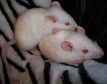
There are differences, however, in the terms used in human medicine and zoology: In mammals, the term albino is often used exclusively for animals with OCA1, in which no residual tyrosinase function is retained and which therefore have white fur, pink skin and red eyes. This is confusing because in humans, in addition to all mutations of the tyrosinase gene, all other disorders of melanin synthesis are also referred to as albinism, syndromes that have very different names in animals.
- Oculocutaneous albinism type 1 corresponds to the albino locus in most mammals.
- Oculocutaneous albinism type 2 corresponds in mammals to the pink eye series ( pink eye dilution P).
- Oculocutaneous albinism type 3 is usually called the brown locus (B) in mammals .
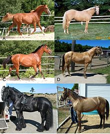
- Oculocutaneous albinism type 4 does not have a uniform name. In horses, for example, the gene is called the cream gene .
- Ocular albinism type 1 .
The individual known mutations are also discussed there.
A number of different genes are referred to as the dilute gene , which lead to a lightening of the color and are usually caused by genes from the albinism spectrum:
- The myosin 5a gene leads to lightened coat color and symptoms related to Griscelly syndrome.
- MLPH is a gene that, like myosin 5a, plays a role in the transport of melanosomes. It tends to lighten the coat color for very similar reasons; some mutations in the gene also cause hair loss.
- The gene for the horse's coat color Champagne is a mutation of the SLC36A1 gene (Solute Carrier 36 family A1), which is also called PAT1 (proton / amino acid transporter 1) or LYAAT1 (lysosomal amino acid transporter 1). It belongs to the same gene family as the gene that causes type 4 oculocutaneous albinism.
- A mutation in the SLC45A5 gene is responsible for the mouse mutation known as golden (gol) dilution .
The silver locus also influences color synthesis, with the result that melanocytes die prematurely, and also leads to numbness and deformities of the eyes in several affected species. Overall, the symptoms are more similar to leucism , although it is a variant of albinism.
In animals, too, there are other as yet unidentified genes that lead to partial or complete albinism.
There are diseases comparable to Chédiak-Higashi syndrome (CHS) and Hermansky-Pudlak syndrome (HPS) in mice, Aleutian mink, Persian cats, cattle, pigs, rats, foxes and even killer whales.
Canine cyclic hematopoiesis also resembles these syndromes. Affected animals are silver-gray, the number of neutrophilic granulocytes is periodically reduced, as are those of red blood cells ( erythrocytopenia ) and platelets ( thrombocytopenia ). This leads to a blood clotting disorder and increased susceptibility to infections. Most of the time the animals die shortly after birth.
Albinism in birds
In addition to melanins, carotenoids and feather structures also play a role in the development of colors in birds .
As in mammals, black and brown colors are produced in birds by eumelanin and pheomelanin. The same genes as in mammals can therefore lead to white or lightened colors in birds.
Carotenoids are ingested with food and lead to red, orange and yellow tones. Birds in which carotenoids are involved in the development of color include the yellow wagtail ( Motacilla flava ), the fitis ( Phylloscopus trochilus ), the blue tit ( Cyanistes caeruleus ), the great tit (Parus major) and the golden oriole ( Oriolus oriolus ). In contrast, the red breast of the robin ( Erithacus rubecula ) is caused by pheomelanin. If the food does not contain enough carotenes, the corresponding feather areas will be white after the next moult. Mutations that lead to disturbances in the accumulation of carotenoids in the feathers are rare.
Both iridescent and non-iridescent blue and green colors are mostly created in birds by the structure of the melanosomes. The melanin is regularly arranged in rods, leaflets, tubes or other structures. The thickness and arrangement of the layers selectively intensifies the visible light color according to the principle of interference on thin platelets . If melanin is missing due to albinism, the structure of the arrangement still selectively intensifies the same color, but the other wavelengths are not filtered out by melanin, so that the bird still appears white overall, sometimes a green or blue shimmer is perceptible where it is otherwise blue or would be green.
Disturbances in dye synthesis in fish, amphibians and reptiles
In fish, amphibians and reptiles, the color of the skin and scales is caused by light interacting with three different types of chromatophores (pigment cells, dye-forming cells): the melanophores, xanthophores and iridophores.
These three cell types, which affect the color of skin and scales, are related to one another. This is why there are some mutations that prevent the production of all three dyes. In addition, there are mutations that prevent dye production in melanophores and xanthophores, but do not affect iridophores. Conversely, there are also mutations that prevent dye production in iridophores and xanthophores, but do not affect melanophores. There are also mutations that affect only one of the three types of dye.
Amelanism
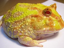
The melanophores correspond in their function to the melanocytes of mammals. They contain melanosomes with melanin as a dye. In most cases, only one type of melanin is synthesized, namely eumelanin. Amelanism is the name given to melanin formation disorders. Sometimes amelanism is mistakenly equated with albinism, but albinism is not limited to amelanism, it also means the absence of pigments other than melanin. Since melanin has essentially the same synthesis path in all animals, mutations in the genes corresponding to the albinism loci of mammals also lead to amelanism in fish, amphibians and reptiles.
Deficiency of iridophore dyes
The iridophores contain reflecting platelets that produce purines. The crystallized purines evoke different color impressions through reflection, often green, blue or an iridescent glitter. They are responsible for the silvery sheen or metallic sheen in fish.
Axanthism
The xanthophores contain pterinosomes, which contain pteridines and flavins as well as carotenes ingested from food. All of these dyes are responsible for yellowish or red colors. If these dyes fail, only that part of the color that is produced by melanins and purines is retained. This state is called axanthism.
Examples
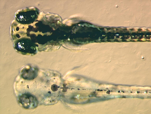
In the zebrafish , larvae with the “sandy” mutation, which cannot produce any melanin, are completely blind for unknown reasons, although the lens and retina appear to be normally developed apart from the lack of melanin. The adult fish are, as to be expected with complete albinism, severely visually impaired and show a pronounced photophobia. Other mutations with the names Golden, Albino, Brass and Mustard also lead to different degrees of albinism, but their larvae are not blind.
Since in fish all fibers of the optic nerves cross over to the other side and not just a part as in mammals, the optic nerve crossing cannot be influenced by melanin.
The leopard gecko ( Eublepharis macularius ) has three mutations that arise independently of one another, which mean that no or less melanin is produced (amelanism). In English they are called "Tremper albino", "Rainwater albino" and "Bell albino". If you cross animals with one of these mutations with animals that have another of these mutations, normal-colored offspring will result.
Syndromes in fish, amphibians, and reptiles associated with albinism
The zebrafish mutant "bleached blond" has a mutation in a gene that produces the Ac45 part of ATP synthase . As embryos, apart from their lightened color, they appear completely normal. Most larvae with this mutation do not develop a swim bladder as they grow further and do not thrive properly, even if they can survive for a few days.
Disorders of dye synthesis in arthropods
Melanin in arthropods
Even with many arthropods ( Arthropoda ) melanin contributes to color. Melanin also plays a role in hardening the cuticle (the hard outer skin) of many arthropods and in their unspecific immune defense against various microorganisms. However, overproduction of melanins is fatal for the animals themselves, so melanin production has to be controlled very precisely.
Insect eye color: ommatochromes and pteridines
The ommatochromes are dyes that appear in the eyes of insects, which include xanthommatin , ommine, and ommidine . In some groups, additional functions of these dyes have evolved, such as coloring the outer shell and excreting tryptophan. The function of the ommatochromes in the coloring of butterfly wings goes back to a single mutation in the family of the noble butterfly (Nymphalidae).
As in the xanthophores of amphibians, reptiles and fish, various pteridines occur in the insect's eye. In the case of the black-bellied fruit fly ( Drosophila melanogaster ) these are, for example, Isoxanthopterin, Pterin, Biopterin, Sepiapterin and Drosopterin. Their biosynthesis begins with the conversion of guanosine triphosphate into dihydroneopterin and then splits up into various synthetic pathways for the individual pteridines.
Many different mutations affect the eye color of insects by preventing their synthesis or by preventing the transport of the various precursors for pteridines or ommatochromes.
Eye colors of the black-bellied fruit fly ( Drosophila melanogaster )
The White, Brown and Scarlet genes from the black-bellied fruit fly ( Drosophila melanogaster ), also known as the fruit fly, encode proteins that belong to the ABC transporters . The gene products of the White, Brown gene work together to produce a membrane protein that transports guanine across the cell membrane. White and Scarlet work together to create a tryptophan transporter.
There are five alleles of the white gene that lead to lightened eye color. In two (w (crr) (H298N) and w (101) (G243S)) the amount of red ( pteridine ) and brown ( xanthommatin ) pigments is equally very much reduced, since both transporters are equally affected. With another mutation (w (Et87)) the lightening of both colors is only slight. In the case of two other mutations (w (cf) (G589E) and w (sat) (F590G)), the production of red pigments is particularly affected, as the mutation mainly affects the effectiveness of the guanine transporter.
The scarlet gene mainly influences the amount of xanthommatin produced . Some of the mutations lead to temperature-dependent dye production, as is known in mammals for the Colorpoint mutations.
Social consequences of albinism for animals
Many social animals also exclude conspecifics that look different or behave unusually as we know it from humans.
The greater tame of the animals associated with albinism or leucism offers a considerable survival advantage in captivity: It makes it easier for people to develop a relationship with the animals. Albino mice bring their young back to the nest more often and more reliably.
Animals with albinism are often crowd favorites in zoos . Albino animals are also extremely popular for animal experiments , because the pigmentless skin is supposedly better suited for this.
selection

A lack of camouflage , impaired vision and higher sensitivity to light can be selection disadvantages caused by albinism. In mice, a significantly reduced mileage and less activity in open terrain were found.

The situation is completely different in captivity: Since the animals are protected by their owners and provided with food, the white color is irrelevant for survival.
In animals that spend their entire life in caves, such as the blind cave tetra ( Astyanax mexicanus ), the camouflage color no longer has a function, since there is no light in which the color could be perceived. White cave shapes are now known from over 80 species of fish.
In addition, white coat color also has a selection advantage. Dark fur emits more polarized light than white, and since insects are able to differentiate polarized and unpolarized light and are attracted by polarized light, white animals are less annoyed by horseflies than dark ones . They can therefore eat more undisturbed and have a lower risk of being attacked by insect-borne diseases.
Xanthism and albinism are not clearly defined

The following terms are not clearly defined:
- Xanthism : yellow or orange animals. Depending on the natural color of the species, either melanins and puridines must be missing in the skin or only one of both. In addition, oculocutaneous albinism type 3 (OCA 3) in humans was also previously referred to as xanthism.
- Albinism has three different meanings:
- only melanin fails completely or partially,
- a white animal due to complete failure of all colorants,
- a lightened to white animal due to complete or partial loss of one or more of the following pigments: melanins, pteridines, carotenes, purines or ommatochromes (partial or complete albinism). This ambiguity does not occur in mammals, since in them only one of the possible dyes contributes to the color of skin, hair and eyes, namely melanin.
- Partial albinism, partial albinism: The word albinism is still used incorrectly in the form of “partial albinism” or “partial albinism” for the animal concerned, often with piebald animals. Genetically speaking, this phenomenon is partial or partial leucism .
literature
- Aleksandra Lipka: Albinism: Search for mutations in the TRP-1 gene. Dissertation, University of Lübeck 2004.
- PM Lund: Oculocutaneous albinism in southern Africa: Population structure, health and genetic care. In: Annals of Human Biology. Volume 32, Number 2, March / April 2005, pp. 168-173.
- Barbara Käsmann-Kellner, Thorsten Schäfer, Christof M. Krick, Klaus W. Ruprecht, Wolfgang Reith, Bernd Ludwig Schmitz: Anatomical differences of the optic nerves, the chiasm and the optic tract in normal and hypopigmented people: a standardized MRI and fMRI -Examination. In: Clinical Monthly Ophthalmology. 220, 2003, pp. 334-344.
- Charlotte Jaeger, Barrie Jay: X-linked ocular albinism. In: Human Genetics. Volume 56, Number 3, pp. 299-304, February 1981.
- Birgit Lorenz, Markus Preising, Ulf Kretschmann: Molecular and clinical ophthalmogenetics. In: Deutsches Ärzteblatt . 98, issue 51-52 of December 24, 2001, pp. A-3445, B-2902, C-2698
- TYRP1 tyrosinase-related protein 1 [Homo sapiens]. NCBI, GeneID: 7306, updated 07-Aug-2007, http://www.ncbi.nlm.nih.gov/sites/entrez?Db=gene&Cmd=ShowDetailView&TermToSearch=7306&ordinalpos=1&itool=EntrezSystem2.PEntrez.Gene.Gene_Results_RVanel
- Markus Kaufmann: Albinism: The tyrosinase gene in 78 variations. Inaugural dissertation . University of Lübeck, 2004.
- Regine Witkowski, Otto Prokop , Eva Ullrich, G Thiel: Lexicon of syndromes and malformations: causes, genetics, risks. Springer, 2003, ISBN 3-540-44305-3 , pp. 86-88.
Web links
- NOAH Albinism Self-Help Group e. V.
- Report: hunting for albinos . Time campus
- Esdras Ndikumana: Gangs of murderers hunt down albinos. Spiegel Online , October 25, 2008.
Individual evidence
- ^ Friedrich Kluge, Elmar Seebold: Etymological dictionary of the German language. Walter de Gruyter, 2002, ISBN 3-11-017473-1 .
- ^ DB van Dorp: Albinism, or the NOACH syndrome (the book of Enoch cv 1-20). In: Clinical genetics. Volume 31, No. 4, April 1987, pp. 228-242, PMID 3109790 .
- ↑ Spanish Piel de Luna means "moon skin". Mexican Association for Albinos, see the following website and Facebook page , (Spanish; accessed July 8, 2019)
- ↑ K. Grønskov, J. Ek, K. Brondum-Nielsen: Oculocutaneous albinism. Orphanet J Rare Dis. 2007 Nov 2; 2:43. PMID 17980020 , PMC 2211462 (free full text)
- ↑ Albinism, Oculocutaneous, Type II; OCA2. In: Online Mendelian Inheritance in Man . (English).
- ↑ David L. Duffy, Grant W. Montgomery, Wei Chen, Zhen Zhen Zhao, Lien Le, Michael R. James, Nicholas K. Hayward, Nicholas G. Martin, Richard A. Sturm: A Three-Single-Nucleotide Polymorphism Haplotype in Intron 1 of OCA2 Explains Most Human Eye-Color Variation. In: Am J Hum Genet. 2007 February; 80 (2), pp. 241-252. PMID 18252222 .
- ↑ SN Shekar, DL Duffy, T. Frudakis, RA Sturm, ZZ Zhao, GW Montgomery, NG Martin: Linkage and association analysis of spectrophotometrically quantified hair color in Australian adolescents: the effect of OCA2 and HERC2. In: Journal of Investigative Dermatology . 2008; Volume 128 (12), pp. 2807-2814. PMID 18528436
- ↑ M. Soejima, H. Tachida, T. Ishida, A. Sano, Y. Koda: Evidence for recent positive selection at the human AIM1 locus in a European population. In: Mol Biol Evol. 2006 Jan; 23 (1), pp. 179-188. Epub 2005 Sep 14. PMID 16162863 .
- ↑ a b c W. Haase: Amblyopia - differential diagnosis. In: Herbert Kaufmann u. a. (Ed.): Strabismus. Enke, Stuttgart 1986, ISBN 3-432-95391-7 , p. 246.
- ↑ a b c d e f g Tony Gamble, Jodi L. Aherns, Virginia Card: Tyrosinase Activity in the Skin of Three Strains of Albino Gecko (Eublepharis macularius) . (PDF; 767 kB) In: Gekko. 5, pp. 39-44.
- ↑ a b Shivendra Kishore and Stefan Stamm: The snoRNA HBII-52 Regulates Alternative Splicing of the Serotonin Receptor 2C. Science 13 January 2006: Vol. 311. no. 5758, pp. 230-232, doi: 10.1126 / science.1118265 .
- ↑ a b c d B. Schüle, M. Albalwi, E. Northrop, DI Francis, M. Rowell, HR Slater, RJ Gardner, U. Francke: Molecular breakpoint cloning and gene expression studies of a novel translocation t (4; 15 ) (q27; q11.2) associated with Prader-Willi syndrome. In: BMC Med Genet. (2005) 6, p. 18.
- ^ A b R. Saadeh, EC Lisi, DA Batista, I. McIntosh, JE Hoover-Fong: Albinism and developmental delay: the need to test for 15q11-q13 deletion. In: Pediatr Neurol. 2007 Oct; 37 (4), pp. 299-302. PMID 17903679
- ↑ C. Fridman, N. Hosomi, MC Varela, AH Souza, K. Fukai, CP Koiffmann: Angelman syndrome associated with oculocutaneous albinism due to an intragenic deletion of the P gene. In: Am J Med Genet A. 2003 Jun 1; 119A (2), pp. 180-183, PMID 12749060 .
- ↑ ANGELMAN SYNDROME; AS. In: Online Mendelian Inheritance in Man . (English).
- ↑ SV Dindot, BA Antalffy, MB Bhattacharjee, AL Beaudet: The Angelman syndrome ubiquitin ligase localizes to the synapse and nucleus, and maternal deficiency results in abnormal dendritic spine morphology. In: Hum Mol Genet . 2008 Jan 1; 17 (1), pp. 111-118. Epub 2007 Oct 16, PMID 17940072 .
- ↑ a b HERMANSKY-PUDLAK SYNDROME; HPS. In: Online Mendelian Inheritance in Man . (English).
- ↑ NCBI Entrez Gene HPS1 Hermansky-Pudlak syndrome 1 (Homo sapiens) GeneID: 3257
- ↑ a b HPS2 Hermansky-Pudlak syndrome 2. In: Online Mendelian Inheritance in Man . (English).
- ↑ HPS3 Hermansky-Pudlak syndrome 3. In: Online Mendelian Inheritance in Man . (English).
- ↑ HPS4 Hermansky-Pudlak syndrome 4. In: Online Mendelian Inheritance in Man . (English).
- ↑ HPS5 Hermansky-Pudlak syndrome 5. In: Online Mendelian Inheritance in Man . (English).
- ↑ HPS6 Hermansky-Pudlak syndrome 6. In: Online Mendelian Inheritance in Man . (English).
- ↑ HPS7 resin Hermansky-Pudlak syndrome 7. In: Online Mendelian Inheritance in Man . (English).
- ↑ HPS8 Hermansky-Pudlak syndrome 8. In: Online Mendelian Inheritance in Man . (English).
- ↑ a b c d e f g h Griscelli Syndrome Type 1 (GS1). In: Online Mendelian Inheritance in Man . (English).
- ↑ a b Griscelli Syndrome Type 2 (GS2). In: Online Mendelian Inheritance in Man . (English).
- ↑ a b Griscelli Syndrome Type 3 (GS3). In: Online Mendelian Inheritance in Man . (English).
- ↑ a b MYOSIN VA; MYO5A. In: Online Mendelian Inheritance in Man . (English).
- ↑ a b RAS-Associated Protein RAB27A. In: Online Mendelian Inheritance in Man . (English).
- ↑ a b melanophilin; MLPH. In: Online Mendelian Inheritance in Man . (English).
- ↑ P. Habermehl, S. Althoff, M. Knuf, J.-H. Höpner: Griscelli syndrome: a case report. In: Clinical Pediatrics. 215, 2003, pp. 82-85.
- ↑ Noah S. Scheinfeld: syndromic albinism: A review of genetics and phenotypes. In: Dermatology Online Journal. 9 (5), p. 5. PMID 14996378
- ↑ a b Lysosomal Trafficking Regulator; LYST. In: Online Mendelian Inheritance in Man . (English).
- ↑ Cockroach . In: Universal Lexicon of the Present and Past . 4., reworked. and greatly increased edition, Volume 9: Johannes – Lackenbach , Eigenverlag, Altenburg 1860, p. 227 .
- ↑ Fried. Ph. Blandin: Albinoism . In: Universal Lexicon of Practical Medicine and Surgery . Leipzig 1835, Volume I, pp. 251–253, here p. 251: “According to the travelers, they [people suffering from albinoism] […] are called Chacrelak in Java. This name is an expression of contempt and describes a type of cockroach or mite that usually hang around in the dark. "
- ↑ John Mason Good: The Study of Medicine . Leipzig 1840, Volume IV, p. 577 [Art. Epichrosis Alphosis, Albinohaut, pp. 576-581]: “In this way, as a result of the discomfort they suffered from the light, and their habit of avoiding it, those who were found on the island of Java were abandoned by the Dutch the contemptuous naming of cockroaches, insects that run around in the dark, is included. "
- ↑ Irenäus Eibl-Eibesfeldt : The biology of human behavior. Piper, Munich / Zurich 1986, pp. 409–417 (Mobbing: “Preservation of group identity”).
- ↑ Martin Franke: Sick and Discriminated. Under the hot African sun, people with albinism have a hard and short life, in: FAS No. 2, January 14, 2018, p. 20.
- ↑ Rico Czerwinski: The hunted. In Tanzania, albinos are tracked like animals and processed into medicine, in: Das Magazin, September 12, 2008. dasmagazin.ch ( Memento from November 23, 2010 in the Internet Archive )
- ↑ Senegal: Albinos face perilous social rejection. In: IRIN News.
- ↑ Tanzania: Search for a missing albino child. on: ORF.at , January 18, 2015.
- ↑ Tanzania: No childhood for albinos arte.tv , ARTE Reportage, 2018, arte.de, September 13, 2018.
- ^ Albinos hit by Zimbabwe's race divide. on: BBC News.
- ↑ Zimbabwe chooses the prettiest albino beauty
- ↑ Jeffrey Gettleman: Albinos, Long Shunned, Face Threat in Tanzania . In: The New York Times . June 8, 2008.
- ↑ J. Chung, J. Diaz: Africans With Albinism Hunted: Limbs Sold on Tanzania's Black Market. August 26, 2010. Retrieved November 29, 2010 .
- ↑ Resolution of the General Assembly, adopted on December 18, 2014. (pdf; 30.6 kB) 69/170. International Albinism Awareness Day. In: un.org. United Nations (UN), February 12, 2015, accessed June 13, 2020 .
- ^ Violence against albinos in Malawi: "Wave of brutal attacks" . Spiegel Online, June 7, 2016.
- ^ John Chiti NJ: Press Articles - Albinism Foundation of Zambia - AFZ. AFZ Albinism Foundation of Zambia, accessed July 19, 2020 (American English).
- ↑ Marcel Safier: Nineteenth Century Images of Albinism - Rudolph Lucasie and family ( Memento of October 30, 2012 in the Internet Archive )
- ↑ a b c d e f g h i Krista Siebel: Analysis of genetic variants of loci for the coat color and their relationships to the color phenotype and to quantitative performance characteristics in pigs. Dissertation . Institute for Livestock Sciences at the Humboldt University of Berlin, July 2001, Chapter 2 (summary of the current state of research).
- ↑ Petra Keller: Investigations into the development of the early acoustic evoked potentials (FAEP) in cats for use in basic research and for clinical application. Dissertation. University of Veterinary Medicine Hannover, 1997.
- ↑ Denis Mariat, Sead Taourit, Gérard Guérin: A mutation in the MATP gene causes the cream coat color in the horse. In: Genetics Selection Evolution. 35, 2003 doi: 10.1051 / gse: 2002039 , pp. 119-133.
- ^ A b D. Cook, S. Brooks, R. Bellone, E. Bailey: Missense Mutation in Exon 2 of SLC36A1 Responsible for Champagne Dilution in Horses. In: PLoS Genet. 2008 Sep 19; 4 (9), p. E1000195. PMID 18802473 .
- ↑ a b c d e f Hein van Grouw: Not every white bird is an albino: sense and nonsense about color aberrations in birds . (PDF; 458 kB) In: Dutch Birding , vol. 28, no. 2, 2006, pp. 79-89.
- ^ H. Durrer, W. Villiger: Schillerradien des Goldkuckucks (Chrysococcyx cupreus (Shaw)) in the electron microscope. In: Cell and Tissue Research. Volume 109, Number 3 / September 1970.
- ↑ H. Durrer, W. Villiger: Schiller colors of the Trogonids. In: Journal of Ornithology. Volume 107, Number 1 / January 1966, doi: 10.1007 / BF01671870 .
- ↑ Matthew D. Shawkey, Geoffrey E. Hill: Significance of a basal layer melanin to production of non-iridescent structural plumage color: evidence from at amelanotic Steller's jay (Cyanocitta stelleri). In: The Journal of Experimental Biology. 209, pp. 1245-1250, doi: 10.1242 / jeb.02115 .
- ↑ a b c d e f Jörg Odenthal, Karin Rossnagel, Pascal Haffter, Robert N. Kelsh, Elisabeth Vogelsang, Michael Brand, Fredericus JM van Eeden, Makoto Furutani-Seiki, Michael Granato, Matthias Hammerschmidt, Carl-Philipp Heisenberg, Yun- Jin Jiang, Donald A. Kane, Mary C. Mullins, Christiane Nüsslein-Volhard: Mutations affecting xanthophore pigmentation in the zebrafish, Danio rerio. In: Development. Vol 123, Issue 1, C 1996, pp 391-398.
- ↑ a b S. K. Frost-Mason, KA Mason: What insights into vertebrate pigmentation has the axolotl model system provided? In: Int J Dev Biol. , 1996 Aug; 40 (4), pp. 685-693. PMID 8877441 .
- ^ Albinism - Biology-Online Dictionary. Retrieved June 29, 2017 .
- ↑ Akihiko Koga, Hidehito Inagaki, Yoshitaka Bessho, Hiroshi Hori: Insertion of a novel transposable element in the tyrosinase gene is responsible for an albino mutation in the medaka fish, Oryzias latipes. In: Molecular and General Genetics MGG. Volume 249, Number 4 / July, 1995, pp. 400-405, doi: 10.1007 / BF00287101 , ISSN 0026-8925
- ↑ A. Koga, Y. Wakamatsu, J. Kurosawa, H. Hori: Oculocutaneous albinism in the i6 mutant of the medaka fish is associated with a deletion in the tyrosinase gene. In: Pigment Cell Res. 1999 Aug; 12 (4), pp. 252-258. PMID 10454293
- ↑ A. Koga, H. Hori: Albinism due to transposable element insertion in fish. In: Pigment Cell Res. 1997 Dec; 10 (6), pp. 377-381. PMID 9428004 .
- ↑ A. Iida, H. Inagaki, M. Suzuki, Y. Wakamatsu, H. Hori, A. Koga: The tyrosinase gene of the i (b) albino mutant of the medaka fish carries a transposable element insertion in the promoter region. In: Pigment Cell Res. 2004 Apr; 17 (2), pp. 158-164. PMID 15016305 .
- ^ ME Protas, C. Hersey, D. Kochanek, Y. Zhou, H. Wilkens, WR Jeffery, LI Zon, R. Borowsky, Clifford J. Tabin : Genetic analysis of cavefish reveals molecular convergence in the evolution of albinism. In: Nat Genet . 2006 Jan; 38 (1), pp. 107-111. Epub 2005 Dec 11. PMID 16341223 .
- ↑ a b c S. K. Frost, LG Epp, SJ Robinson: The pigmentary system of developing axolotls.
- ^ Stephan CF Neuhauss, Oliver Biehlmaier, Mathias W. Seeliger, Tilak Das, Konrad Kohler, William A. Harris, Herwig Baier: Genetic Disorders of Vision Revealed by a Behavioral Screen of 400 Essential Loci in Zebrafish. In: The Journal of Neuroscience. October 1, 1999, 19 (19), pp. 8603-8615.
- ↑ Adam Amsterdam, Shawn Burgess, Gregory Golling, Wenbiao Chen, Zhaoxia Sun, Karen Townsend, Sarah Farrington, Maryann Haldi, Nancy Hopkins: A large-scale insertional mutagenesis screen in zebrafish. In: Genes & Development. 1999. 13, pp. 2713-2724.
- ↑ M. Zhao, I. Söderhäll, JW Park, YG Ma, T. Osaki, NC Ha, CF Wu, K. Söderhäll, BL Lee: A novel 43-kDa protein as a negative regulatory component of phenoloxidase-induced melanin synthesis. In: J Biol Chem . 2005 Jul 1; 280 (26), pp. 24744-24751. Epub 2005 Apr 27. PMID 15857824
- ↑ K. Söderhäll, L. Cerenius: Role of the prophenoloxidase-activating system in invertebrate immunity. 1: In: Curr Opin Immunol. 1998 Feb; 10 (1), pp. 23-28. PMID 9523106 .
- ↑ RD Reed, LM Nagy: Evolutionary redeployment of a biosynthetic module: expression of eye pigment genes vermilion, cinnabar, and white in butterfly wing development. In: Evol Dev. 2005 Jul-Aug; 7 (4), pp. 301-311. PMID 15982367 .
- ^ A b c Alfred M. Handler, Anthony A. James: Insect Transgenesis: Methods and Applications. CRC-Press, 2000, ISBN 0-8493-2028-3 , pp. 81ff.
- ↑ a b S. M. Mackenzie, MR Brooker, TR Gill, GB Cox, AJ Howells, GD Ewart: Mutations in the white gene of Drosophila melanogaster affecting ABC transporters that determine eye coloration. In: Biochim Biophys Acta . 1999 Jul 15; 1419 (2), pp. 173-185. PMID 10407069 .
- ↑ AJ Howells: Isolation and biochemical analysis of a temperature-sensitive scarlet eye color mutant of Drosophila melanogaster. In: S. Biochem Genet. 1979 Feb; 17 (1-2), pp. 149-158. PMID 110313 .
- ↑ Vitus B. Dröscher : White lions must die. Rules of the game of power in the animal kingdom. Rasch and Röhring, Hamburg 1989, pp. 212–244 (Mobbing: "Kill the outsider!").
- ↑ M. Protas, M. Conrad, JB Gross, C. Tabin, R. Borowsky: Regressive evolution in the Mexican cave tetra, Astyanax mexicanus. In: Current biology: CB. Volume 17, Number 5, March 2007, pp. 452-454, ISSN 0960-9822 . doi: 10.1016 / j.cub.2007.01.051 . PMID 17306543 . PMC 2570642 (free full text).
- ^ R. Borowsky, H. Wilkens: Mapping a Cave Fish Genome: Polygenic Systems and Regressive Evolution. In: J Hered. 2002 Jan-Feb; 93 (1), pp. 19-21. PMID 12011170 .
- ↑ Gábor Horváth1, Miklós Blahó, György Kriska, Ramón Hegedüs, Balázs Gerics, Róbert Farkas, Susanne Åkesson: An unexpected advantage of whiteness in horses: the most horsefly-proof horse has a depolarizing white coat. In: Proc. R. Soc. B. June 7, 2010, vol. 277 no. 1688, pp. 1643-1650, doi: 10.1098 / rspb.2009.2202 .
- ↑ M. Tsudzuki, Y. Nakane, N. Wakasugi, M. Mizutani: Allelism of panda and dotted white plumage genes in Japanese quail. In: J Hered. 1993 May-Jun; 84 (3), pp. 225-229. PMID 8228175 .
- ↑ M. Miwa, M. Inoue-Murayama, H. Aoki, T. Kunisada, T. Hiragaki, M. Mizutani, S. Ito: Endothelin receptor B2 (EDNRB2) is associated with the panda plumage color mutation in Japanese quail. In: Anim Genet. , 2007 Apr; 38 (2), pp. 103-108. Epub 2007 Feb 22. PMID 17313575 .
