
Leucism


Leucism (from ancient Gr. Λευκός leukós "white") is a defect mutation in animals, which leads to the fact that the fur is white and the skin underneath is pink, since the skin does not contain any melanocytes (dye-forming cells). In contrast, in albinism, the cells are present but unable to produce the pigment melanin .
Genes whose mutation leads to leucism were previously commonly abbreviated with "W". In early embryonic development, they cause the structures of the neural crest to develop incorrectly , which means that no or very few melanoblasts migrate out of the neural crest. This means that there are no longer any pigment-forming cells on the body surface . Areas that are directly related to the central nervous system , especially the eyes , usually have at least a certain number of pigment-forming cells, so that the eyes of leucistic animals are colored light brown or dark blue to orange (in snakes dark blue (almost black) to blue), depending on how many pigment cells there are. There are leucistic genes that cause health impairments, especially when they are homozygous , but also those that do not cause impairments; the combination with other genes is sometimes important here.
Most forms of spotting can be traced back to weakened forms of leucism.
It is now known that there are several different genes whose mutations can lead to leucism. These include the endothelin receptor B gene (EDNRB), the paired box gene 3 (PAX3), SOX10, the microphthalmia-associated transcription factor (MITF), c-kit and the steel locus (encoding MGF).
Mutations of individual gene locations
Endothelin receptor B gene
The endothelin receptor B gene (EDNRB) encodes a G-protein coupled receptor , which is produced in various cell types and is responsible for the differentiation of melanocytes and nerve cells of the intestine, the neural folds before the total closure of the neural tube and the complete formation of the neural crest .
Mutations in the endothelin receptor B gene therefore lead to spotting or leucism (pink skin and white fur) and often an overstretching of the large intestine ( megacolon ) due to missing nerve cells in the intestine ( aganglionosis ). The megacolon leads to death within a few hours to days after birth.
People (EDNRB)
A small number of cases of Hirschsprung's disease can be traced back to mutations in the endothelin receptor.
A mutation of the endothelin receptor also leads to Waardenburg-Shah syndrome or WS4.
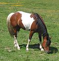
Lethal white syndrome in horses (OLWS)

Heterozygous carriers of a mutation of the endothelin receptor in horses almost always have a frame-over-spotting caused by this gene. Foals homozygous for the frame overo gene are completely white and die within 24 hours of intestinal obstruction due to the lack of intestinal innervation. This is called Lethal-White Syndrome.
- See also Overo-Lethal-White-Gene
Rats (EDNRB)

Heterozygous animals with the spotting lethal (s l ) gene, which is a mutation of the endothelin receptor, are pied. The colored spots are on the head, tail and form a black stripe on the back. Other than that, they are healthy.
Rats homozygous for this gene (s l s l ) are completely white with dark eyes. Sometimes they have small spots on the head and hips. They develop a megacolon due to a lack of nerves and usually die from it within the first week of life.
Japanese quail ( Coturnix japonica ) (EDNRB)
In the Japanese quail ( Coturnix japonica ), one of the two variants of the EDNRB gene in birds, namely EDNRB2, is responsible for the colors panda and dotted white.
c kit
c-Kit is a transmembrane receptor tyrosine kinase from the family of platelet growth factor ( PDGF ) and CSF-1 (colony stimulating factor-1) receptors.
c-Kit is effective as a proto-oncogene in malignant melanoma. C-Kit expression has been demonstrated in some solid tumors. Defects in c-Kit lead to myeloid leukemia.
The c-Kit receptor is involved in the reproduction, differentiation, functional maturation and maintenance of a large number of differentiated cells. Its mutations can cause anemia and sterility. They are also responsible for various forms of leucism and spotting.
Human (c-kit)

Piebaldism - white spots on the skin, usually combined with a white forelock - is also caused in humans by the KIT locus.
Mouse (c-kit)

The mouse c-kit locus is known under the following names, among others: c-kit, belly-spot, dominant spotting, spotted sterile male, steel factor receptor, dominant white spotting, proto-oncogene protein
Most of the mutations in this locus are incompletely dominant.
Homozygotes or mice with two different c-Kit mutations on the alleles are usually dark-eyed and completely white. Depending on the severity of the mutation, it is associated with other health problems such as sterility, liver damage and anemia to varying degrees. Many die in the womb or shortly after birth. They can only survive if the residual function of the c-Kit is sufficient. The belly-spot mutant, even homozygous, only has a white spot under the belly and is otherwise healthy.
Heterozygous mice are viable and are spotted to varying degrees, particularly on the abdomen and tail.

Horses (c-kit)
The kit locus is on chromosome 3 of the horse (ECA3).
One of several forms of Sabino check is caused by a mutation of the c-KIT locus.
The same applies to the Roan gene, which in heterozygous form leads to spiky-haired horses with mixed white and colored hair. The roan gene is homozygous.
It has also been proven for the Tobiano check that it is caused either by the kit locus itself or by a gene location that is in the immediate vicinity on the same chromosome.
It has also been shown that the horse's dominant white color can also be traced back to this locus.
 Tobiano checkCommons : Tobiano - collection of images, videos and audio files
Significant Sabino check

Minimal sabino: If the gene is simply present, it leads to clear markings on the face and legs with at most small spots on the stomach.

Permanent red mold: Genetically not a mold, but a fox with white prickly hair


Beef (c-kit)
The white face of the Hereford cattle is caused by the c-Kit locus, and due to this mutation there is an increased susceptibility to a rapidly growing cancer of the nictitating membrane (3rd eyelid). The white face of the Simmental cows can be traced back to another mutation, the genetic location of which has not yet been determined. It is very likely that the typical spotting known from Holstein and Holstein cattle can also be traced back to the kit locus.

Holstein cows

Hereford bull


Pig (c-kit)
Both the pink color of most high-performance breeds and various check patterns as well as the belt markings of some pig breeds are caused by mutations in the c-Kit locus.
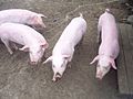
Norwegian country pig
The pink color of the pigs is caused by a mutation of the c-Kit locus.
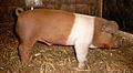
Belt drawing - also a c-kit mutation


Cat (c-kit)
The Birman cat's white gloves are caused by a mutation of the c-Kit locus. The light-colored body fur and blue eyes, on the other hand, are the result of partial albinism , since melanocytes are present here, but due to a modified enzyme they can only produce melanin in the cooler parts of the body.
The genes that are responsible for the white spots in normal domestic cats have not yet been identified (2011).

Birman cat with white gloves, caused by a mutation in the KIT locus

Pied cats have their spots from a mutation of c-Kit
.jpg)

Pax3
The Pax3 gene (paired box gene 3, also paired domain gene 3 or paired box homeotic gene 3) is involved in controlling early embryonic development as a transcription factor .
It belongs to the group of Pax genes , which typically have a paired box domain and a paired type homeodomain.
Slight mutations of this locus lead to different forms of spotting and leucism. Other mutations cause deafness. There are severe malformations of the brain and nervous system in which the spine often does not close until birth ( spina bifida ) or the skull remains open. The circulatory system can also be severely affected, with many of the main arteries starting in the wrong place, veins that normally recede being preserved, and the like. Severe mutations in the gene therefore lead to death before birth.
Human (Pax3)
In humans, mutations of the gene cause Waardenburg syndrome type 1 and 3 (WS1 and WS3), craniofacial-deafness-hand syndrome (literally: facial skull-deafness-hand syndrome) and rhabdomyosarcoma .
Mouse (Pax3)
Most of the knowledge about the Pax3 gene was obtained through mutations in the mouse.
In addition to mutations that lead to death in the womb or shortly after birth due to severe malformations of the nervous and circulatory system, there are also animals that are only spotted and otherwise healthy.
Common features include white feet, a kink in the tail, an open neural tube, various malformations, and dislocations of the main arteries.
Microphthalmia-Associated Transcription Factor (MITF)
The microphthalmia-associated transcription factor (MITF) is a protein from the group of transcription factors . Mutations in his gene can lead to various forms of leucism (spotting). In addition, malformations of the eyes ( microphthalmia ) occur. In contrast to most forms of leucism, animals that are leucistic or piebald due to a mutation of this locus often have red or pink eyes rather than blue or brown.
Human ( MITF )
In humans, the MITF gene is on chromosome 3 ( GeneID 4286 ) and causes Tietz syndrome and Waardenburg syndrome type 2 (WS2). In addition, the activity of the MITF gene in metastatic melanoma is detected in 10-16 % of all cases. The exact role of MITF in the development of melanoma and in its progression is still unclear.
Fruit fly (Mitf)
Mutations of this gene also lead to malformations of the eyes in fruit flies (Drosophila melanogaster) , which supports the theory that compound eye and lens eye did not develop independently of one another.
Mouse (Mitf)

Mutations in the mouse by this gene range from animals that are white, light-colored, or piebald but otherwise healthy and have light-colored, red or pink eyes, to animals with severe malformations or with a complete lack of eye and bone compaction to varying degrees . There are also animals that are deaf.
Sox10
Sox 10 is a transcription factor that is necessary for the formation of the neural crest in embryonic development. It is necessary for the development of melanocytes and oligodendrocytes , which isolate the axons of the nerve cells from one another in the central nervous system . It also plays a key role in the development of the peripheral and intestinal nervous systems.
Human (Sox10)
Mutations of the Sox10 locus cause Waardenburg syndrome type 4, the Yemenite deaf-blind hypopigmentation syndrome and the neurological variant of Waardenburg-Shah syndrome, which combines the megacolon of Hirschsprung's disease with the symptoms of Waardenburg syndrome.
More severe mutations mean that the child dies in the womb during embryonic development.
Mouse (Sox10)
In addition to various mutations that lead to leucism or spotting without further signs of disease, there are also mutations in mice with a megacolon, which cause the young to die shortly after birth.
Steel locus
The Steel-Locus (Steel factor, SF, Steel) works closely with the c-Kit-Locus and is therefore also called Kitl (Kitlg, Kit ligand). Further names and abbreviations for the steel locus are: Sl; SLF; contrasted (Con), cloud gray (Clo), grizzle-belly (Gb), stem cell factor (SCF), mast cell growth factor (Mgf).
Like all leucism loci, it leads to leucism in various forms.
The steel factor plays an essential role in the development of the cells of the germ line into finished egg and sperm cells and in their migration to the later reproductive organs. Therefore, many mutations in this locus lead to infertility. Other common consequences of mutations in this locus are excessive bone density and anemia due to decreased blood formation. Since steel also influences the development of mast cells, mutations can lead to disorders of the immune system. There is also deafness.
Mouse (steel)
Much lightened mutations lead to animals with white fur and normal colored eyes. Slightly lightened animals are either uniformly gray or have lighter areas or white spots on their ears, feet, tip of the tail and on the belly.
In addition to animals that are healthy in every respect apart from their light fur, there are some mutations that are associated with infertility and anemia, or in which homozygous animals die while still in the womb.
Beef (steel)
The color of mixed white and dark hair of the white-blue Belgian and the Shorthorn cattle is caused by a mutation of the steel locus. In homozygous animals, the mutation leads to completely white fur and sterility of some of the cows (white heifer disease), as the uterus is not fully developed.

White-blue Belgian homozygous for the mutation of the steel locus
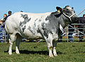
White-blue Belgian heterozygous for the mutation of the steel locus
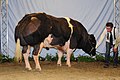
White-blue Belgian without this mutation


See also
- Albinism
- Coat color
- Coat colors of dogs
- Coat colors of the cat
- Coat colors of horses
- Genetics of horse colors
- Domestic rabbit genetics
- Melanism
swell
Endothelin receptor
- Ling Hou, William J. Pavan, Myung K. Shin and Heinz Arnheiter: Cell-autonomous and cell non-autonomous signaling through endothelin receptor B during melanocyte development. In: Development. 2004, 131, pp. 3239-3247 Published by The Company of Biologists doi: 10.1242 / dev.01193
- Cheryl E. Gariepy, Daniel T. Cass, Masashi Yanagisawa: Medical Sciences Null mutation of endothelin receptor type B gene in spotting lethal rats causes aganglionic megacolon and white coat color. In: Proc. Natl. Acad. Sci. USA . Vol. 93, pp. 867-872, January 1996, http://www.pnas.org/cgi/content/abstract/93/2/867?ck=nck
- DL Metallinos, AT Bowling, J. Rine: A missense mutation in the endothelin-B receptor gene is associated with Lethal White Foal Syndrome: an equine version of Hirschsprung disease. In: Mamm Genome. 1998 Jun; 9 (6), pp. 426-431. PMID 9585428 .
- EM Santschi, PD Vrotsos, AK Purdy, JR Mickelson: Incidence of the endothelin receptor B mutation that causes lethal white foal syndrome in white-patterned horses. In: Am J Vet Res. 2001 Jan; 62 (1), pp. 97-103. PMID 11197568
- GC Yang, D. Croaker, AL Zhang, P. Manglick, T. Cartmill, D. Cass: A dinucleotide mutation in the endothelin-B receptor gene is associated with lethal white foal syndrome (LWFS); a horse variant of Hirschsprung disease (HSCR). In: Human Molecular Genetics . 1998, Vol. 7, No. 6, pp. 1047-1052.
- Jeanne Amiel, Tania Attié, Dominique Jan, Anna Pelet, Patrick Edery, Christelle Bidaud, Didier Lacombe, Paul Tam, Juliette Simeoni, Elisabeth Flori, Claire Nihoul-Fékété, Arnold Munnich, Stanislas Lyonnet: Heterozygous endothelin receptor B (EDNRB) mutations in isolated deer jump disease. In: Human Molecular Genetics. 1996 Vol. 5, No. 3, pp. 355-357.
- ↑ M. Tsudzuki, Y. Nakane, N. Wakasugi, M. Mizutani: Allelism of panda and dotted white plumage genes in Japanese quail. In: J Hered. 1993 May-Jun; 84 (3), pp. 225-229. PMID 8228175
- ↑ M. Miwa, M. Inoue-Murayama, H. Aoki, T. Kunisada, T. Hiragaki, M. Mizutani, S. Ito: Endothelin receptor B2 (EDNRB2) is associated with the panda plumage color mutation in Japanese quail. In: Anim Genet. 2007 Apr; 38 (2), pp. 103-108. Epub 2007 Feb 22. PMID 17313575
c kit
- NCBI - Kit kit oncogene (Mus musculus)
- Olga Maksimovic: Studies on the expression of the c-kit receptor in the development and progression of malignant melanoma. Inaugural dissertation to obtain a doctorate in medicine, University Dermatology Clinic Tübingen
- Edwin N. Geissler, Melanie A. Ryan, David E. Housman: The dominant-white spotting (W) locus of the mouse encodes the c-kit proto-oncogene. In: Cell. Volume 55, Issue 1, October 7, 1988, pp. 185-192, doi : 10.1016 / 0092-8674 (88) 90020-7
- RA Fleischman: Human piebald trait resulting from a dominant negative mutant allele of the c-kit membrane receptor gene. In: J Clin Invest. 1992 Jun; 89 (6), pp. 1713-1717. PMID 1376329
- SA Brooks, E. Bailey: Exon skipping in the KIT gene causes a Sabino spotting pattern in horses. In: Mamm Genome. 2005 Nov; 16 (11), pp. 893-902. Epub 2005 Nov 11. PMID 16284805
- Hai-Bin Ruan, Nian Zhang, Xiang Gao: Identification of a Novel Point Mutation of Mouse Proto-Oncogene c-kit Through N-Ethyl-N-nitrosourea Mutagenesis. In: Genetics. 2005 Feb; 169 (2), pp. 819-831. PMID 15731517
- M. Johansson Moller, R. Chaudhary, E. Hellmén, B. Höyheim, B. Chowdhary, L. Andersson: Pigs with the dominant white coat color phenotype carry a duplication of the KIT gene encoding the mast / stem cell growth factor receptor In : Mammalian Genomes . Springer, New York, ISSN 0938-8990 , Volume 7, Number 11 / November 1996, doi: 10.1007 / s003359900244 , pp. 822-830.
- E. Giuffra, G. Evans, A. Tornsten, R. Wales, A. Day, H. Looft, G. Plastow, L. Andersson: The Belt mutation in pigs is an allele at the Dominant white (I / KIT) locus . In: Mamm Genome. 1999 Dec; 10 (12), pp. 1132-1136. PMID 10594235
- E. Giuffra, A. Tornsten, S. Marklund, E. Bongcam-Rudloff, P. Chardon, JM Kijas, SI Anderson, AL Archibald, L. Andersson: A large duplication associated with dominant white color in pigs originated by homologous recombination between LINE elements flanking KIT. In: Mamm Genome. 2002 Oct; 13 (10), pp. 569-577. PMID 12420135
- MP Cooper, N. Fretwell, SJ Bailey, LA Lyons: White spotting in the domestic cat (Felis catus) maps near KIT on feline chromosome B1. In: Anim Genet. 2006 April; 37 (2), pp. 163-165. doi: 10.1111 / j.1365-2052.2005.01389.x .
- S. Marklund, M. Moller, K. Sandberg, L. Andersson: Close association between sequence polymorphism in the KIT gene and the roan coat color in horses. In: Mamm Genome. 1999 Mar; 10 (3), pp. 283-288. PMID 10051325
- C. Mau, PA Poncet, B. Bucher, G. Stranzinger, S. Rieder: Genetic mapping of dominant white (W), a homozygous lethal condition in the horse (Equus caballus). In: Journal of Animal Breeding and Genetics. 121 (6), Volume 121, Issue 6, pp. 374-383 doi: 10.1111 / j.1439-0388.2004.00481.x .
- N. Reinsch, H. Thomsen, N. Xu, M. Brink, C. Looft, E. Kalm, GA Brockmann, S. Grupe, C. Kuhn, M. Schwerin, B. Leyhe, S. Hiendleder, G. Erhardt , I. Medjugorac, I. Russ, M. Forster, R. Reents, G. Averdunk: A QTL for the degree of spotting in cattle shows synteny with the KIT locus on chromosome 6. In: J Hered. 1999 Nov-Dec; 90 (6), pp. 629-634. PMID 10589513
- MD Grosz, MD MacNeil: The "spotted" locus maps to bovine chromosome 6 in a Hereford-Cross population. In: J Hered. 1999 Jan-Feb; 90 (1), pp. 233-236. PMID 9987932
- Sheila Schmutz: Genetics of Coat Color Patterns in Cattle. Whiteface. Status: Feb. 17, 2004,
- Sheila M. Schmutz: Cat Coat Color Genetics. As of October 18, 2011,
- L. Magnol, MC Chevallier, V. Nalesso, S. Retif, H. Fuchs, M. Klempt, P. Pereira, M. Riottot, S. Andrzejewski, BT Doan, JJ Panthier, A. Puech, JC Beloeil, MH de Angelis, Y. Herault: KIT is required for hepatic function during mouse post-natal development. In: BMC Dev Biol. 2007 Jul 5; 7, p. 81. PMID 17612398
- MC Penedo, LV Millon, D. Bernoco, E. Bailey, M. Binns, G. Cholewinski, N. Ellis, J. Flynn, B. Gralak, A. Guthrie, T. Hasegawa, G. Lindgren, LA Lyons, KH Roed, JE Swinburne, T. Tozaki: International Equine Gene Mapping Workshop Report: a comprehensive linkage map constructed with data from new markers and by merging four mapping resources. In: Cytogenet Genome Res. 2005; 111 (1), pp. 5-15. PMID 16093715
Pax3
- Waardenburg syndrome, type I. In: Online Mendelian Inheritance in Man . (English).
- Craniofacial-deafness-hand syndrome. In: Online Mendelian Inheritance in Man . (English).
- Rhabdomyosarcoma 2. In: Online Mendelian Inheritance in Man . (English).
- Waardenburg syndrome, type III. In: Online Mendelian Inheritance in Man . (English).
- Pax3 paired box gene 3 (Mus musculus)
- M. Ptok, S. Morlot: Unilateral inner ear hearing loss due to mutations in the PAX3 gene in Waardenburg syndrome type I. In: ENT. Springer, Berlin / Heidelberg, Volume 54, Number 7 / July 2006, ISSN 0017-6192 , pp. 557-560. doi: 10.1007 / s00106-005-1315-1
MITF
- NCBI: Mitf (Drosophila melanogaster) , GeneID: 3885647, as of 21-Feb-2007
- NCBI: Mitf microphthalmia-associated transcription factor (Mus musculus) , GeneID: 4286, status: 05-Apr-2007
- Phenotypic Alleles ( Memento from September 27, 2011 in the Internet Archive )
- NCBI: MITF microphthalmia-associated transcription factor (Homo sapiens) , GeneID: 4286, as of 08-Apr-2007
- NCBI: Tietz syndrome, MIM: 103500
- NCBI: Waardenburg syndrome, type IIA, MIM: 193510
- NCBI: Waardenburg syndrome / ocular albinism, digenic, MIM: 103470
Sox10
- Sandra Wißmüller: Biochemical analysis of the properties of the HMG domain of the transcription factor Sox10 . Natural science faculties of the Friedrich-Alexander-Universität Erlangen-Nürnberg, 2006.
- Michael Wegner: Mechanistic analysis of the interaction between the transcription factors Sox10 and Pax3. Chair of Biochemistry and Pathobiochemistry, Institute of Biochemistry, University of Erlangen.
- NCBI: Sox10 SRY-box containing gene 10 (Mus musculus) , GeneID: 20665, as of: 24-Mar-2007
- NCBI: SOX10 SRY (sex determining region Y) -box 10 (Homo sapiens) , GeneID: 6663, status: 08-Apr-2007
Steel
- NCBI: Kitl kit ligand (Mus musculus). GeneID: 17311, as of 05-Apr-2007
- Mouse Genome Informatics: Kitl. Phenotypic Alleles as of April 16, 2007
- C. Charlier, B. Denys, JI Belanche, W. Coppieters, L. Grobet, M. Mni, J. Womack, R. Hanset, M. Georges: Microsatellite mapping of the bovine roan locus: a major determinant of White Heifer disease . In: Mamm Genome. 1996 Feb; 7 (2), pp. 138-142. PMID 8835531
- JJ Seitz, SM Schmutz, TD Thue, FC Buchanan: A missense mutation in the bovine MGF gene is associated with the roan phenotype in Belgian Blue and Shorthorn cattle. In: Mamm Genome. 1999 Jul; 10 (7), pp. 710-712. PMID 10384045
- Sheila Schmutz: Genetics of Coat Color Patterns in Cattle. The Roan Pattern. Status: February 17, 2004