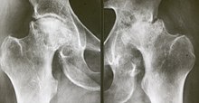
Femoral head necrosis
| Classification according to ICD-10 | |
|---|---|
| M87.05 | Idiopathic aseptic bone necrosis: pelvic region and thigh [pelvis, femur, buttocks, hips, hip joint, sacroiliac joint] |
| ICD-10 online (WHO version 2019) | |
The femoral head necrosis (actually incorrectly: femoral head necrosis ) is a disease that is characterized by the death of part of the bony head of the femur . The disease is one of the aseptic bone necrosis . The cause is decreased blood flow, which leads to necrosis .
A special case of femoral head necrosis is Perthes' disease in childhood. The causes of the circulatory disturbance, which is also present here, are still unknown and are being discussed controversially.
causes
In adults, the exact causes have not been fully clarified; femoral head necrosis occurs more frequently in diabetes mellitus , alcoholism and after high-dose (mostly systemic) cortisone treatment. Long-term treatment with anticoagulants can also result in bone necrosis. Post-traumatic femoral head necrosis can occur after injuries to the femoral head.
Clinical picture

For no apparent cause, a hip begins to hurt. The mobility of the joint is restricted; in most cases internal rotation and extension are inhibited. The normal X-ray image often cannot show any pathological changes in the first stage; only the examination with the MRI shows the changes in the metabolic situation in the diseased bone in the early stage. If the disease progresses, remodeling processes appear in the still living part of the bone, and the body tries to seal off the dead part. The necrotic part of the femoral head finally collapses, the joint can then hardly be loaded.
Coxarthrosis destructive rapide (CDR) differs from this clinical picture , which is accompanied by rapid destruction of the femoral head and the socket within a few months with clinically and diagnostically identical symptoms.
Femoral head necrosis, on the other hand, is usually a process that takes years and is often only recognized late.
The image is an MRI representation of a right-sided femoral head necrosis (to the left of the viewer). A sharply demarcated area of the bone is swollen edematously , the boundary layer is particularly noticeable, here the body deals with the disease process. The left hip joint is normal, this can be seen by simple comparison.
therapy
In the past, attempts were mostly made with little success to remove the dead part of the bone, to reline it with cancellous bone and thus to achieve a stable femoral head again. Furthermore, the vascular pedicled bone graft implantation is still a sensible alternative to joint preservation in younger patients when there are still fewer signs of femoral head necrosis in the MRI and in the X-ray examination. This can be useful, especially for younger patients, if this connection is stable. The leg is then less flexible, but it can withstand unrestricted loads. In the meantime, the surgical use of an artificial hip joint ( endoprosthesis ) has become the method of choice to restore the patient's leg to a load. The arthrodesis , ie the operative stiffening of the hip joint is practically no longer used today.
In smaller dogs (<15 kg body mass), surgical removal of the dead femoral head often brings about sufficient clinical improvement. A “false joint” is formed , which allows the animal sufficient movement and is largely free of pain.
In off-label use , prostacyclin and prostacyclin analogues such as B. Ilomedin or Iloprost , used for the treatment of bone marrow edema and femoral head necrosis.
literature
- S3- guideline atraumatic femoral head necrosis in adults of the German Society for Orthopedics and Orthopedic Surgery (DGOOC). In: AWMF online (as of December 31, 2013)
Individual evidence
- ↑ KL Krämer, M Stock, M. Winter: Clinic Guide Orthopedics . Jungjohann Verlag Stuttgart 1991, ISBN 3-8243-1139-9 .
- ↑ Petje et al .: Aseptic bone necrosis in childhood . In: Orthopäde , 10, 2002, pp. 1027-1038. doi: 10.1007 / s00132-004-0634-3
- ↑ Thorsten Schmidt: Infusion, femoral head drilling or infusion after femoral head drilling in the treatment of atraumatic femoral head necrosis (FKN) and bone marrow edema syndrome . (PDF; 1.3 MB; 76 pages) Chair for Orthopedics at the Clinic of the University of Regensburg, dissertation, 2009:
