Cortex and cancellous bone
Substantia corticalis (short cortex , from Latin cortex 'cortex' ) and substantia spongiosa (short spongiosa , from Latin spongia 'sponge' ) are the two macroscopic forms of bone tissue in the bone .
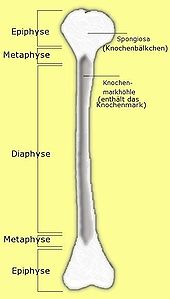
- The cortex consists of a compact layer of bone tissue just below the periosteum , which forms the surface of the bone. The shaft of long bones the cortex is very thick and is therefore also called the substantia compacta (short Kompakta from Latin compactus , compact ' refers).
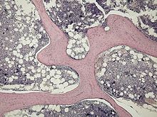
- The cancellous bone as an internal bone structure, on the other hand, lies inside the bone; The bone tissue is organized here as a spongy system of fine trabeculae , in whose cavities the bone marrow is located. In the flat bones , the cancellous bone is called Diploë , the blood vessels in it are called Venae diploicae (also Breschet veins in the skullcap ).
Construction principle
The cancellous bone forms a closely-meshed framework, with most of the trabeculae being arranged along the most important stress lines ( tension trajectories ) of the bone. The architecture depends on whether the section of the bone is mainly exposed to pressure , such as the vertebral bodies , or bending and torsional forces , such as the femoral head . This lightweight construction principle enables the saving of bone substance with sufficiently high stability and thus a lower weight of the bone.
There is no cancellous bone in the shaft of long bones, instead the cortex is particularly pronounced. Since the greatest compression and expansion occurs at the edge when it is bent, this distribution of the bone tissue also corresponds to the lightweight construction.
histology
Both the cortex and the cancellous bone consist of lamellar bone in the mature bone . But while the lamellae in the cortex are organized in osteons , that is, concentrically surrounded by Haversian canals and supplied by the vessels running in them, the lamellae of the cancellous bone run largely parallel to the trabecular surface. The trabeculae are vascular so that the osteocytes in them have to be nourished by diffusion from the vessels of the bone marrow, which limits the thickness of the trabeculae to 300 µm as a rule.
Genesis of the cancellous bone
In desmal ossifying bones, the primary cancellous bone arises from the fusion of ossification points; the resulting primary trabeculae consist of woven bones . The primary cancellous bone of the chondral ossifying bones is created by chondroclasts leaving small bars of mineralized cartilage, which are then covered by osteoblasts with a layer of woven bone . In both cases the primary trabeculae are within the scope of remodeling in secondary trabeculae converted from lamellar bone.
modification
The remodeling mechanisms in the cortex and cancellous bone differ: While the cortex is remodeled by building new osteons, remodeling in the cancellous bone is done using Howship lacunae . The result of the remodeling process gives the bone density . The turnover rate is much higher in the cancellous bone, so that diseases with loss of bone substance (e.g. osteoporosis ) initially lead to the loss of cancellous bone, which manifests itself in fractures of the femoral neck and vertebral bodies , where the cancellous bone is of particular importance.
literature
- Benninghoff, Drenckhahn: Anatomie Vol . 1 . 17th edition. Elsevier, Urban & FischerVerlag, Munich 2008, ISBN 978-3-437-42342-0 , pp. 133-136, 274-276 .
- Keith L. Moore, T. Vidhya N. Persaud: Embryology . 5th edition. Elsevier, Urban & FischerVerlag, Munich 2007, ISBN 978-3-437-41112-0 , pp. 424-426 .
Individual evidence
- ↑ Barbara I. Tshisuaka: Breschet, Gilbert. In: Werner E. Gerabek , Bernhard D. Haage, Gundolf Keil , Wolfgang Wegner (eds.): Enzyklopädie Medizingeschichte. De Gruyter, Berlin / New York 2005, ISBN 3-11-015714-4 , pp. 208 f.
- ↑ Renate Lüllmann-Rauch: Pocket textbook histology . 5th edition. Thieme, Stuttgart 2015, ISBN 978-3-13-129245-2 , p. 169 .


