Hoffa-Kastert syndrome
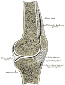
As Hoffa Kastert syndrome is a hypertrophy of the corpus adiposum infrapatellar ( Hoff Ash fatty substance called). It is a complex of symptoms resulting from mechanical or inflammatory changes in the knee joint .
| Classification according to ICD-10 | |
|---|---|
| M79.4 | Hypertrophy of the infrapatellar corpus adiposum (Hoffa-Kastert syndrome) |
| ICD-10 online (WHO version 2019) | |
history
When the orthopedic surgeon Albert Hoffa described the inflammatory hypertrophy of the Hoffaschen fat body (Corpus adiposum infrapatellare) in 1904 , he assumed that it was a disease in its own right, a disease sui generis, i.e. of its own kind. It was only decades later (1953) that the surgeon Josef Kastert discovered that the changes in the fat body were almost always associated with internal damage to the knee joint. These include meniscus damage , free joint bodies or cartilage disorders . The hypertrophy of the Hoffaschen fat pad is not an independent disease. It is a symptom of primary internal knee damage. The new name Hoffa-Kastert syndrome is more appropriate to the facts. The name Hoffasche disease has medical and historical significance.
Hypertrophy of the Hoffaschen fat pad
causes
Internal knee damage can be caused by traumatic effects on the knee joint, which occur directly, indirectly, once or repeatedly.
complaints
Spontaneously or after an injury to the knee joint, increasing pain occurs in the joint, which is often associated with restricted mobility. Joint swelling is not always present. When the knee is bent, a feeling of tension occurs. A joint blockage caused by the enlarged Hoffasche fat body is softer and less fixed than with a meniscus entrapment.
Finding
Fat body hypertrophy manifests itself as pain on both sides of the inside and outside of the knee joint. A bulge-like swelling can be observed on both sides of the patellar ligament . The examiner feels a soft to firm thickening that is painful to pressure at these points. When pressure is applied on both sides of the patellar ligament, the kneecap can rise without there being any joint effusion.
Pathological-anatomical changes
The inflammatory co-reacting Hofasche fat body enlarges and takes on a coarse consistency. It is yellowish-reddish in color or soaked in blood. The microscopic examination of the fat body shows round-cell immigration into the area around the vessels and an increase in connective tissue cells .
Diagnosis
First and foremost is the clinical examination. MRI and knee arthroscopy should also be mentioned as further diagnostic measures . If hypertrophic villi of the fat body are trapped, a bloody joint effusion can occur.
therapy
In most cases, the diagnosed internal knee damage can only be treated surgically. Partial resections on the Hofaschen fat body are possible in exceptional cases if villi have caused entrapment or if the overview needs to be improved during the anterior cruciate ligament surgery . If the primary damage in the joint has been successfully repaired, the hypertrophy of the Hoffaschen fat pad regresses without further treatment.
literature
- Hartmut Zippel: Meniscus injuries and damage . Verlag Johann Ambrosius Barth, Leipzig 1973, p. 203.
- Wilfried Wehner, Eberhard Sander (ed.): Trauma surgery . VEB Verlag Volk und Gesundheit, Berlin 1981, p. 206.
- Gerhard Kaiser: Guide to Orthopedics . 2nd Edition. VEB Gustav Fischer Verlag, Jena 1963, p. 212.
- Hamilton Bailey: Surgical Medical Examination. 6th edition. Verlag Johann Ambrosius Barth, Leipzig 1974, p. 543.
- Renate Jäckle ( edit .): HEXAL LEXICON Orthopedic Rheumatology . Verlag Urban & Schwarzenberg, Munich / Vienna / Baltimore 1992, p. 121.
Individual evidence
- ^ Josef Kastert: The adhesions of the fat body of the knee joint as an independent clinical picture . In: The surgeon . tape 9 . Springer, Berlin Heidelberg 1953, p. 390 .
