Kneecap
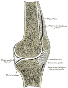
The kneecap ( Latin patella ) is a flat, disc-shaped, triangular bone when viewed from the front, which lies in front of the knee joint , in whose articular surfaces it is involved. The kneecap acts as a sesamoid in the tendon of the quadriceps femoris muscle ( four-headed thigh muscle ). It protects the knee joint and multiplies the strength development of the quadriceps by extending the lever arm. The ossification of the kneecap occurs in humans via several ossification centers from the age of three.
anatomy
The kneecap has two surfaces:
- The facies anterior ("front surface") is convex ( arched outwards ) and has small openings through which the supplying vessels pull into the interior of the bone. Superficial portions of the quadriceps tendon cover the kneecap and thus continue from the quadriceps distal (remote from the body) into the patellar ligament (kneecap ligament). Most of the quadriceps tendon radiates into the kneecap from proximally (close to the body) and exits distally to form the patellar ligament. Further ahead, one ensures bursa that prepatellar bursa for upholstery and displaceability to the skin.
- The upper two thirds of the posterior face ("back surface") is covered by hyaline articular cartilage , which, at around 6 mm, is the thickest articular cartilage in the human body. The posterior face has a vertical ridge that fits into the gap ( intercondylar sulcus ) between the condyles ( articular knots ) of the femur . It divides the posterior surface into two facets, which in turn are connected (articulate) with the condyles. Usually the angle between the two facets is 120 ° -140 °. The lateral ( side ) facet is usually much wider. According to Wiberg and Baumgartl, four patella shapes are distinguished depending on the relationship between the facets .
The kneecap has three edges corresponding to the two surfaces:
- The upper, strong edge ( Margo superior ) serves as an attachment (insertion surface) for the quadriceps, more precisely for two of its muscle heads, the rectus femoris and vastus intermedius muscles .
- The inner edge ( Margo medialis ) tapers away from the body and serves as the attachment surface for the medial part of the quadriceps ( Musculus vastus medialis ).
- The lateral edge ( Margo lateralis ) also tapers away from the body and serves as the attachment surface for the lateral part of the quadriceps ( Musculus vastus lateralis ).
The tip that tapers downwards ( apex patellae ) serves as the origin for the ligamentum patellae, which extends to the shin .
Special features in birds
The kneecap of birds behaves essentially like that of mammals . It is relatively large in water fowl. Due to the particularities in the anatomy of the quadriceps muscles in birds are nomenclaturally as Musculi femorotibiales referred. The kneecap of the birds has an additional muscle groove ( sulcus musculi ambientis ) on the front .
function
The kneecap increases the distance between the force vector of the quadriceps and the center of rotation of the knee joint and thereby extends the lever arm of the extensor muscles of the thigh. The result is a lever extension, which results in a power saving of up to 44%. The patella centralizes the forces generated by the four heads of the quadriceps femoris muscle and transfers them distally to the patellar ligament and tibial tuberosity with little friction. The forces acting on the quadriceps attachment tendon and the patellar ligament are different. The patella takes on a protective function for the femur and increases the contact area with it. It ensures a better distribution of the force on the femur and offers the advantage of two joint surfaces covered with articular cartilage. During the stretching movement ( extension ), the kneecap covers a distance of about eight to ten centimeters above the thigh bone.
Due to the high forces in the kneecap joint that can occur during stress, the joint to which the patella belongs is the joint most frequently affected by osteoarthritis in people in Germany.
Diseases of the kneecap
Malformations of the kneecap can be in the form of a missing ( aplasia ) or insufficient formation ( patellar dysplasia ), a lack of fusion of the bone nuclei ("split kneecap", patella partita ) or an inadequate formation of the plain bearing with the possibility of lateral jumping out, a dislocation of the kneecap ( Patellar luxation ) occur.
An external force can lead to a fracture of the kneecap ( patellar fracture ).
Inflammatory changes at the base of the kneecap ligament can lead to the tip of the kneecap becoming detached ( patellar tip syndrome , Larsen-Johansson disease ). If the contact pressure of the kneecap is disturbed in its slide bearing, the cartilage coating on the back of the kneecap is insufficiently supplied and thus cartilage damage ( chondropathia patellae ). Chronic changes in the kneecap joint ( osteoarthritis ) usually manifest themselves behind the kneecap ( retropatellar arthrosis ).
Changes in the context of syndromes
Typically, changes in the patella occur in the following diseases:
- Diastrophic dysplasia
- Meier-Gorlin Syndrome
- Popliteal pterygium syndrome all possibly agenesis
- Ehlers-Danlos Syndrome
- Ellis van Creveld syndrome
- Osteogenesis imperfecta
- Marfan's Syndrome
- Rubinstein-Taybi Syndrome
- Trisomy 21 all possible dislocation
- Nail-patella syndrome hypoplasia
- Aniridia-patellar aplasia syndrome
Individual evidence
- ↑ a b Wolfgang Dihlmann : Joints, vertebral connections: Clinical radiology including computer tomography - diagnosis, differential diagnosis. Georg Thieme Verlag, 2002, ISBN 3-13-132013-3 , p. 335 ff.
- ↑ a b M. Schünke: Functional anatomy: topography and function of the movement system. Thieme, Stuttgart 2000. ISBN 978-3-13-118571-6
- ↑ F.-V. Salomon: Textbook of Poultry Anatomy. Gustav Fischer, Jena 1993, ISBN 3-334-60403-9 .
- ↑ AMJ Bull, AA Amis: Biomechanics. In: D. Kohn (Ed.): Knie. Thieme. Stuttgart 2005.
- ↑ B. Tillmann, W. Peterson: Functional Anatomy. In: D. Kohn (Ed.): Knie. Thieme, Stuttgart 2005.
- ↑ P. Aglietti, PPM Menchetti: Biomechanics of the Patellofemoral Joint. In: GR Scuderi (Ed.): The Patella. Springer Verlag, New York 1995.
- ↑ DT Reilly, M. Martens: Experimental analysis of the quadriceps muscle force and patellofemoral joint reaction force for various activities. In: Acta Orthopedica Scandinavia , 1972, 43, pp. 126-137.
- ^ Health in Germany. Data tables. Federal health reporting. Robert Koch Institute , Berlin 2006.
- ↑ F. Hefti: Pediatric Orthopedics in Practice. Springer 1998, p. 649, ISBN 3-540-61480-X .


