Knee joint
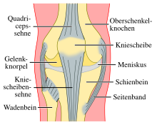
The knee joint ( Latin articulatio genus ) is the largest joint in mammals located in the knee ( Latin genu ) . The thighbone (femur) , the shinbone (tibia) and the kneecap (patella) form the bony joint bodies.
The knee joint is a compound joint. It consists of two individual joints, the kneecap joint (Articulatio femoropatellaris) , which is located between the thighbone and the kneecap, and the throat joint (Articulatio femorotibialis) , which is located between the thighbone and the tibial head (Caput tibiae) . Anatomically seen, it is at the proximal joint between the tibia ( tibia ) and fibula ( fibula ) (Articulatio tibiofibularis) Although a standalone joint, but usually a bulge of the knee joint capsule ( recess subpopliteus ) is connected with the knee joint.
Is located at the back of the knee, the knee (popliteal fossa) , in the depth of important blood vessels and nerves pass. The popliteal lymph nodes (Lymphonodi poplitei) are also formed here.
etymology
The common. Body part name mhd. Knee , ahd. KNEO goes on IE. Ḡenu- "knee" back.
Bony structures and articular surfaces
The joint surfaces of the joint bodies are covered with hyaline joint cartilage , which deforms under the effect of transmitted forces and thus provides the contact surfaces on which the surface pressure creates the necessary balance of forces. The joint surfaces can only transmit compressive forces. Since, as a rule, only parts of the joint surfaces function as contact and bearing surfaces, the actual pressing pressures are significantly higher than the pressures estimated from the anatomical joint surfaces, they exceed the blood pressure many times over. Therefore, there are no blood vessels in the articular cartilage, because the blood pressure could not maintain an exchange of substances against the tissue pressure. The joint space between the two cartilage surfaces is very thin: only a few molecular layers of synovial fluid fit into it. The dimension of the joint space is in the micrometer range and cannot be shown in the X-ray image. What orthopedists refer to as the “joint gap” are the two radiolucent cartilage layers of the joint body. The articular cartilage is traversed by collagen fibers , which on the one hand attach it to the bone and on the other hand give it a certain tensile strength. At the joint space, they pivot in a horizontal direction (the so-called tangential fiber layer) and help the joint bodies slide.
Thigh bone
The femur ends knee Windwärts ( distally ) with two fairly wide, slightly outwardly curved ( convex ) condyle ( lateral femoral condyle and medial femoral condyle ) between which a narrow hole extending from front to back and on the back (intercondylar notch) is . The surface of the articular knots (condyli ossis femoris) is laid out in a spiral shape, so that the center of the rotational movement of the joint describes a spiral movement path under the movement. The inner articular knot is one to two centimeters larger in the horizontal direction ( sagittal ) than the outer one and is further away from the joint.
The cartilage layer of the femur is thinner than that of the kneecap, with the outer sliding surface being covered by a thicker layer of cartilage than the inner one.
The thigh gnarls diverge somewhat further away from the joint and to the rear. The outer articular knot is wider at the front than at the back, while the inner articular knot has a uniform width. In horizontal planes, the articular knots are only slightly curved around a vertical axis. In the vertical plane, the curvature increases towards the rear, that is, the radius of curvature becomes smaller. The inner joint cartilage is also curved around a vertical axis (rotational curvature).
On the articular surface of the thigh bone (front side) there is a flat gliding channel for the kneecap ( facies patellaris femoris or trochlea ossis femoris ) between the two articular knots . This sliding channel divides the joint surfaces into two facets. The outer facet is slightly larger and runs closer to the joints and further forward than the smaller inner one. In contrast to this, it can absorb more pressure, especially during flexion.
Particularities in quadruped mammals
In quadrupedal mammals, the thigh bones have an outward curvature, the radius of which becomes larger towards the rear. As a result, excessive flexion (hyperflexion) results in greater tension in the side ligaments ( collateral ligaments ), which slows down the movement (so-called spiral joint ). The joint is always in a flexed position. In dogs, for example, the maximum extension does not exceed an angle of 150 ° open to the rear.
Special features in birds
The femur is short in birds . It is used to shift the starting point of the leg above the bird's center of gravity in order to enable the most energy-saving possible standing and walking. The center of gravity of most birds is very low, around the level of the knee joint (see also bird skeleton ).
Shin
The upper end of the shinbone also ends in two, slightly inwardly curved articular knots ( condylus lateralis tibiae and condylus medialis tibiae ). In between are a sublime bone First (tibial) , located in two small bumps divided ( tubercle intercondylar media and tubercle intercondylar lateral ) and two trough configurations ( intercondylar area anterior - in animals intercondylar area cranial and - posterior intercondylar area - in animals intercondylar area caudalis ). The entire upper surface of the tibia is called the tibia plateau, which forms the articular surface of the tibia (Facies articularis superior tibiae) for the knee joint. Since the ridge of the bone extends over the entire tibial plateau, the rotational movement is retained as a possible direction of movement of the joint.
Kneecap
The kneecap is triangular and slightly arched outward on its front surface. It is embedded as a sesamoid in the tendon of the four-headed thigh muscle ( quadriceps femoris muscle ) , which embeds it coming from above. The fibers of the patellar ligament (ligamentum patellae) arise from its lower tip (apex patellae) . On the back of the kneecap (facies articularis patellaris) there is a ridge that divides the articular surfaces into two facets. Your cartilage layer is about six millimeters thick.
When the knee is bent, the kneecap lies firmly in the furrow just above the joint space between the thighbone and shinbone, and further above when the leg is extended. Therefore, it can be shifted a little to the right and left in the extended position and relaxed muscles, but not in the flexed position.
The main task of the kneecap is to lengthen the lever arm and thus the torque of the quadriceps, as it increases the distance between its line of action from the center of movement of the knee joint. It also serves to guide the tendon and reduces the resistance to sliding movement of the tendon over the bone.
Special features in dogs
In addition to the kneecap, dogs have three other sesamoid bones on the knee joint. The fabellae are located in the tendon of origin of the two heads of the two-headed calf muscle ( gastrocnemius muscle ) . The side fabella is larger than the central one. Both articulate with the thigh bone. The third sesamoid lies in the tendon of origin of the popliteal muscle ( musculus popliteus ) in the hollow of the knee.
Joints
Kneecap joint
The kneecap joint (articulatio femoropatellaris) is the joint between the thigh bone and the kneecap. The joint surface covered with hyaline cartilage on the back of the kneecap (facies articularis patellae) and that on the front of the thighbone (facies patellaris femoris) face each other. When flexed and stretched, the kneecap slides about five to ten millimeters over the thigh bone in the groove provided for it, the entry into the groove occurs at around 30 ° of flexion. This type of joint is also known as a sledge joint (articulatio delabens) .
Forces in the kneecap joint
The force with which the kneecap acts on the femur is known as the patellofemoral joint reaction force (PFJR). It is the resulting force vector from the vectors of the tendon of the quadriceps femoris muscle and the patellar ligament. The size of the PFJR depends on the angle at which the knee joint is located and the force exerted by the quadriceps femoris muscle. With increasing flexion in the knee joint, the contact surfaces of the joints shift proximally, the total contact surface increases between 20 ° and 90 °. The joint surfaces have their greatest possible contact at around 90 °. With increasing flexion, the contact pressure increases and reaches a maximum at an angle of 70–75 °.
The PFJR can exceed several times your own body weight. When walking it is about half the body weight (0.2–0.4 kg; 0.5 kg), when climbing stairs 3.3 kg and when doing deep squats 7.6 kg. When running, the joint reaction forces can be between 7 and 11.1 kg, 17.5 kg when lifting weights up to 24 times the body weight for a downward jump.
Particularities in quadruped mammals
The kneecap ligament is divided into three parts in horses and cattle . One distinguishes a pointing towards the middle (medial) , a side (lateral) and a middle (intermediate) patellar ligament ( patellar ligament media , patellar ligament lateral and patellar ligament intermedium ). Only the middle one attaches to the tibia, the other two each to the side of it.
For the lateral attachment of the kneecap, two retaining straps ( ligamentum femuropatellare mediale and ligamentum femuropatellare lateral ) are formed between its lateral edges and the thigh bone. In the case of predators ( dogs , cats ) these are only very inconspicuous.
The kneecap joint has another special feature in horses. The knot pointing towards the middle has a clear elevation at the end near the joint, the so-called "nose" (tuberculum trochleae ossis femoris) . The kneecap can be hooked onto this bead with the loop between the inner and middle kneecap ligament. The knee is passively fixed in the extended position - largely without the use of muscle power - which enables almost fatigue-free standing. One leg is fixed in this way, while the other rests relaxed on the tip of the hoof (“shielding”). After a while, the rested leg is fixed and the previously locked leg is completely relieved. By pulling the quadriceps femoris muscle and pulling the biceps femoris muscle sideways , the kneecap is released from this rest position, which restores full mobility of the joint.
Popliteal joint
The knee joint (articulatio femorotibialis) is the actual joint responsible for bending the knee. It is a mixture of a wheel and a hinge joint which is as Drehscharnier-, angle of rotation - ( Trochoginglymus ) or bicondylar joint designated (thus allowing the flexion and extension, as well as bent at a 90 ° knee a slight inward and outward rotation) and is between the outwardly curved thigh gnarls and the tibial plateau. It has to withstand great loads, but at the same time allow sufficient mobility.
Menisci
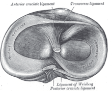
Since the connected (articulating) joint surfaces do not fit exactly, this "inequality" (incongruence) is compensated by crescent-shaped fiber cartilage discs, the menisci , which can follow the rotational movements. Another task of the menisci is to increase the contact area between the tibia and the femur.
A distinction is made between an inner meniscus (meniscus medialis) , which is C-shaped, larger and somewhat more immobile (because it is fused with the inner ligament), and an outer meniscus (meniscus lateralis) , which is circular, smaller and more flexible (as it is not fused with any collateral ligament) ). The menisci are wedge-shaped in cross section. The high edge is on the outside, the low one on the inside. Since the thigh gnarls lie exactly in the middle directly on the tibial plateau and peripherally on the menisci, they carry a substantial part of the load.
When the knee joint is moved, the menisci are pushed forward by the thigh condyles (also: 'thigh knots', derived from "tree stump" or "block of wood", see also: thigh bones ): When flexed, the two condyles roll and slide back and push the menisci backwards, when they stretch they move forward again. When the lower leg is rotated outwards, the outer meniscus is pushed forward on the shinbone, the inner meniscus is withdrawn, and when the lower leg is rotated it is the other way round.
The menisci can be connected at the front by a short, strong ligament ( ligamentum transversum genus ) , but this is variable and has no connection to the tibial plateau. As the actual anchoring of the menisci, the fibers of the front and rear horns radiate into the tibia plateau and thus establish the considerable tensile strength. In addition, variably applied ligaments ( ligamenta meniscofemoralia ) can connect the lateral meniscus with the inner femur.
The inner meniscus can be felt in the joint space towards the middle on the patella ligament. If the inner meniscus is damaged, pressure pain can be triggered here. The following examination procedures, so-called meniscus signs, are also diagnostically helpful :
- Steinmann-I (Steinmann-I symbol) : When the lower leg is bent, the knee is rotated. Pain during internal rotation indicates an injury to the external meniscus, while external rotation indicates an injury to the internal meniscus.
- Steinmann II (Steinmann II sign) : When the knee joint is flexed, the pressure pain moves from the front to the back (because the menisci move backwards when the knee is bent).
- Apley grinding test : rotation with the knee joint flexed in the prone position. Pain analogous to the Steinmann I sign.
- Böhler's sign : pain during abduction or adduction (valgus and varus stress) in the knee joint.
- Payr sign : print on the inside cross-legged. Any pain that occurs indicates an internal meniscus lesion.
Joint capsule, fluid and space
The knee joint is encased in a wide knee joint capsule (capsula articularis genu) . This is strongly tense and stabilized when fully extended. With increasing flexion, it slackens.
Outer layer
The outer layer of the joint capsule (Membrana fibrosa capsulae) is only very stable on the back of the joint, where it attaches to the edge of the femur near the joint and pulls to the pit between the bones. There she advertises in the front indentation of the shin plateau. The joint space enclosed by the two layers has a horseshoe shape in horizontal section. On the side, the outer layer has a gate that serves as a passage for the popliteal muscle ( musculus popliteus ) . Far from the joint, the capsule attaches to the edges of the shinbones and is there firmly attached to the menisci. At the front, the joint cavity is bounded by the kneecap and the kneecap ligament, with which it is also fused.
An important task of the outer layer is the sensitive care of the knee. Receptors embedded in it provide important information about the position and changes in the pull and thus enable cooperation with the muscles of the entire lower extremity.
Inner layer
The inner layer of the joint capsule is called the synovial membrane (Membrana synovialis capsulae) . It rests on the front surface of the thighbone, follows the bone border on the backside and runs there over the pit between the thighbones.
The inner skin forms the joint fluid ( synovia ) that is important for the nutrition of the cartilage . Movements of the knee joint mix the synovial fluid and thereby improve the absorption of nutrients by the cartilage cells ( chondrocytes ) . The right amount and composition of the synovial fluid is also of crucial importance for the lubrication of the knee joint. They minimize the friction of the corresponding cartilage surfaces during the rolling-sliding movement.
Fat body
The knee joint has a fat body ( corpus adiposum infrapatellaris , also called " Hoffasch fat body "), which is located in front of the joint between the two layers of the joint capsule. When flexed, it is squeezed (compressed) by the tensioned ligament under the kneecap and mainly bulges out to the side.
The continuations of the fat body along the sides of the kneecap and the ligament under the kneecap are called fat folds ( plicae alares) . They can be divided into two trains:
- Plica synovialis infrapatellaris , which originally divided the joint into two chambers
- Plica alaris that pulls towards the lateral edges of the kneecap
Bursa
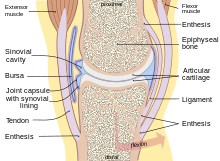
To avoid damage to the pulling via the joint ligaments, the knee joint has at particular points of friction above, before and below the knee joint bursa (bursa) , some of which have a connection to the joint space ( Bursa or recess suprapatellaris and infrapatellar bursa ). The infrapatellar bursa is pushed between the patella ligament and the tibia. A bursa is located in the back of the joint cavity under the attachment tendon of the half-skinned muscle ( musculus semimembranosus ) (bursa musculi semimembranosi) . Two more lie under the tendons of origin of the two-headed calf muscle ( musculus gastrocnemius ) ( bursa subtendinea musculi gastrocnemii lateralis and bursa subtendinea musculi gastrocnemii medialis ).
Two bursa have no connection to the joint space and are therefore self-contained. One lies under the skin in front of the kneecap ( bursa subcutanea prepatellaris ) , the other lies between the kneecap ligament and the outer layer of the joint capsule (bursa infrapatellaris profunda) .
Bulges
In front of the kneecap (suprapatellar) , the joint capsule on the front of the thighbone forms a bulge ( bursa or suprapatellar recess ) that smooths or unfolds when flexed and thus enables the kneecap to move up to seven centimeters. On the sides of the kneecap, there are additional folds of the capsule (recessus parapatellaris) . The subpopliteus recess is located on the posterior side of the joint, under the tendon of origin of the popliteal muscle .
Tapes
Since the knee is very unstable due to its bony construction, it is secured by numerous ligaments . They strengthen the joint capsule, in the outer layer of which they are usually built. The ligaments of the knee are divided into anterior (ventral) , lateral (collateral) , posterior (dorsal) and central ligaments according to their location .
Front tape lock
The five to six millimeters thick patellar ligament ( patellar ligament , "patellar tendon") is part of the capsule and pulls as a continuation of the quadriceps from the lower edge of the patella to the anterior roughening of the tibia, the tibial bump (tibial tuberosity) , where it attaches a large area. Another ligament (retinaculum patellae) runs centrally and to the side of the kneecap and the patella ligament . This is divided into a central portion (retinaculum medial patellar) , the wide from the directed to the middle thigh muscle ( vastus medialis ) is formed and a lateral portion (retinaculum lateral patellar) extending from the lateral wide thigh muscle ( vastus lateralis ) forms . They are part of the outer layer of the joint capsule.
Lateral tape protection
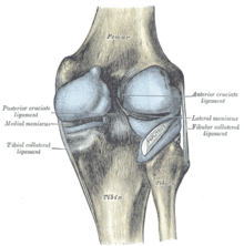
The knee has two collateral ligaments: an inner (ligamentum collaterale tibiale) and an outer (ligamentum collaterale fibulare) . In the extended position, both side ligaments (also known as collateral ligaments ) are taut and thus prevent the rotation, in the flexed position the radius of curvature decreases, the origin and attachment approach each other and the ligaments are relaxed as a result. Both collateral ligaments stabilize the knee joint in a lateral direction ( frontal plane ) so that it is prevented from kinking in a bow-leg position ( genu varum ) or knock-kneel position ( genu valgum ).
The inner ligament ( Ligamentum collaterale tibiale or mediale ) is a triangular, flat ligament that runs over a wide area from the top of the inner thighbone (epicondylus medialis femoris) to the inside of the shin (Facies medialis tibiae) . It is built into the outer layer of the joint capsule and grown together with the inner meniscus.
There are three different fiber groups:
- The front long fibers run from the top of the inner thigh bone to the inside of the shin.
- The posterior upper short fibers radiate into the medial meniscus.
- The posterior lower long fibers pass from the medial meniscus to the shin.
If the inner ligament is torn, the lower leg can be moved to the side (“opening phenomenon”).
The outer ligament ( ligamentum collaterale fibulare or laterale ) is a strong ligament, which in its dorsal section extends in a cylindrical shape from the attachment of the lateral thigh bone (epicondylus lateralis femoris) to the fibula head (caput fibulae) . It has no fixed connection to the joint capsule and the menisci.
The anterolateral ligament described in 2013 extends from the same origin as the outer ligament to the anterolateral tibia and is fused there in the middle between the fibula head and the tibial tuberosity . It is also firmly attached to the outer meniscus.
Rear tape backup
There are two ligaments on the back of the knee joint. The oblique popliteal ligament ( ligamentum popliteum obliquum ) arises at the attachment point of the half-skinned muscle ( musculus semimembranosus ) on the inner tibial gnar and strengthens the posterior side of the joint capsule with which it fuses. The arched popliteal ligament ( ligamentum popliteum arcuatum ), on the other hand, pulls from the posterior fibula over the insertion of the popliteal muscle and also pulls up the capsule in the middle as a reinforcement.
Central tape backup
The cruciate ligaments (ligamenta cruciata) run from the pit between the femur to the tibia. Viewed from the side and from the front, they cross each other in their course.
By preventing the joint surfaces from sliding forward or backward ( translation ), the cruciate ligaments stabilize the knee. In addition, they inhibit the rotational movement, especially the inward rotation, in which they wrap themselves around each other and the anterior cruciate ligament becomes taut. During the outward rotation, they unwind, which means that the knee is always turned a little outward when it is maximally extended ( final rotation ). The classic injury of the anterior cruciate ligament therefore occurs, e.g. B. when skiing , with bent knee and inward turning under the action of force.
The position of the cruciate ligaments in relation to the joint capsule is a specialty. They are located within the outer layer of the joint capsule (intracapsular), but outside the inner skin. This saves the cruciate ligaments in an open, sharp U-shape towards the rear. Thus, they lie outside the actual joint cavity (extra-articular). In terms of evolutionary history, this fact can be explained by the fact that the cruciate ligaments migrated from behind during evolution, pushing the inner capsule skin with them.
In the event of isolated injury to one of the two cruciate ligaments, the “ drawer phenomenon ” occurs: In the event of a complete tear ( rupture ) of the anterior cruciate ligament, the shin bone can be moved further forward than the uninjured knee compared to the thigh bone;
Anterior cruciate ligament
The anterior cruciate ligament ( ligamentum cruciatum anterius - in animals, ligamentum cruciatum craniale ) extends from the anterior depression between the tibial glands to the side and slightly backwards to attach to the inside of the lateral thigh gnar. It is divided into a front-center and a rear-side bundle. Due to the wide diversification of the original area of these bundles, part of the anterior cruciate ligament is taut both when flexed and when stretched. This prevents hyperextension (hyperextension) when the leg is extended, while it counteracts the forward movement of the shin when the leg is flexed (“front drawer”).
Posterior cruciate ligament
The posterior cruciate ligament ( ligamentum cruciatum posterius - in animals ligamentum cruciatum caudale ) is stronger and has its origin in the posterior depression of the tibial plateau and pulls forward-centered to attach to the lateral anterior surface of the inner thigh knot. It tenses when you flex and thus prevents the shin from sliding backwards (rear drawer). When the leg is extended, the posterior cruciate ligament supports the anterior one in preventing hyperextension. However, its main role is to stabilize the knee when it is flexed and under load.
Musculature
The ligaments are supported by the surrounding muscles . Precise execution of movements, especially in the flexed position, can only be carried out through cooperation and changing settings of the ligamentous apparatus and muscles.
Straightener
The front of the thigh bone is surrounded by a large four-headed extensor muscle ( Musculus quadriceps femoris ) . The three broad muscles ( vastus medialis , vastus lateralis and vastus intermedius muscles ) as well as the straight muscle ( rectus femoris muscle ) form these four heads. They straighten the knee by starting at the tibia bulge . The kneecap is embedded between these muscles and their joint attachment, which ends as the patella ligament. The extensor ( extensor ) thus initially attaches to the kneecap. From there, the force is transmitted to the lower leg via the patella ligament.
In birds, the iliotibialis cranialis muscle is the only extensor muscle of the knee joint.
Flexor
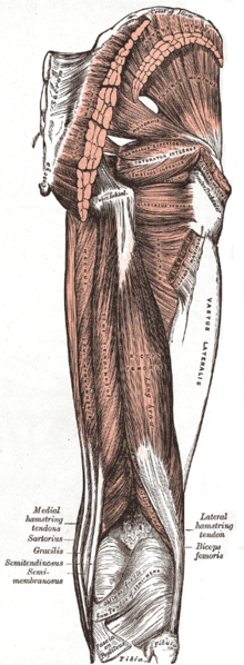
Flexors ( flexor ) of the knee joint is inside the longest muscle of the body, the so-called cutter muscle ( musculus sartorius ) . Together with two other muscles, the slender thigh muscle ( gracilis muscle ) and the half-tendon muscle ( semitendinosus muscle ), it forms a joint attachment further inside the shin, the so-called goosefoot ( pes anserinus superficialis) . Other flexors of the knee joint are the two-headed thigh muscle ( biceps femoris ) and the two-headed calf muscle ( gastrocnemius ) , which is part of the three-headed lower leg muscle ( triceps surae ) .
In birds, the take musculus iliofibularis , muscle ischiofemoralis , muscle iliotibial lateralis , the Musculi femorotibiales , the muscle ambiens , the flexor cruris medialis and the flexor cruris lateralis flexion of the knee joint.
Flexors and Inward Twists
The half-tendon muscle (semitendinosus muscle) , the half-skinned muscle (semimembranosus muscle) , the slender thigh muscle (gracilis muscle) and the popliteal muscle (popliteus muscle) also act as flexors, but are also responsible for inward rotation ( internal rotator ).
Away from home
The outward rotation (external rotation) is done by the biceps femoris muscle.
Movements
The knee joint allows in humans because of its surrounding joint capsule and ligaments located inside and outside the same, only the diffraction ( flexion ) and stretching ( extension ) up to about 150 °. Due to the lack of a pair of joint bodies, there is no local center of movement (such as in the hip joint ). Instead, flexion and extension result in a combination of rolling and sliding movement of the joint body, which is known as a sliding bearing . With maximum extension there is also - with the ligamentous apparatus intact - a secondary movement, the so-called final rotation , in which the shinbone rotates a few degrees outwards.
The knee joint is a so-called swivel-hinge joint ( trochoginglymus ) . It has five degrees of freedom . A distinction is made between three degrees of freedom of movement and two degrees of freedom of rotation. The degrees of freedom of movement mean the front-rear ( anterio-posterior ) and mid-side (medio-lateral) displacement as well as pressure (compression) and tension (traction) . Flexion and extension as well as inward and outward rotation ( rotation ) are defined as degrees of freedom of rotation . However, the rotary movements are only possible in the bent state.
- Flexion up to approx. 120–150 °
- Extension up to approx. 5–10 °
- Inward rotation of 10 ° (at 90 ° flexion)
- Outward rotation by 30–40 ° (at 90 ° flexion)
In domestic dogs , the knee joint is at an angle of approximately 130 ° to 140 ° in the resting position. The total range of motion for flexion and extension is between 90 and 130 ° and can even be exceeded with passive movement. In normal gait, however, the knee only moves between 110 ° in the flexed position and 150 ° in the extended position, so the range of motion is only about 40 °. In the extended position, internal and external rotation are only possible to a very limited extent (5–10 °); in the bent position, the knee can be rotated about 10–20 ° outwards and 20–45 ° inwards.
Arteries
The arterial supply of the knee joint is provided by a large number of different arteries , which anastomose with one another and thus form a dense collateral network ( rete articulare genus ). They include:
- Descending artery genus
- Arteria genus media
- Arteria superior lateralis genus
- Arteria superior medialis genus
- Inferior lateral artery genus
- Arteria inferior medialis genus
Diseases
Fractions and dislocations
After a dislocation ( dislocation ) , the knee joint is seldom fully usable again, as a large number of ligament structures tear in this injury. The popping out of the kneecap is called a patellar dislocation . Congenital knee dislocation is a special form .
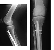
The bone parts that form the knee joint can break . Such breaks (fractures) must be treated surgically using osteosynthesis . The bone parts are fixed with steel or titanium plates and so-called set screws or intramedullary nails (plate osteosynthesis) . Often it is also necessary to straighten the articular surface, which has sunk in the accident, and to reline it with the body's own bone material or ceramic material. Pure cracks can also only be fixed with screws. Damage to the kneecap (kneecap fractures = patellar fractures) is very rare. They are usually the result of a direct fall on the kneecap. The kneecap breaks into several parts. It can longitudinal , transverse or mixed fractures occur. Simple fractures heal with appropriate treatment without permanent damage. Complex rubble fractures usually leave a functional restriction. A transverse hernia must always be treated surgically , otherwise the enormous forces of the quadriceps lead to pseudarthroses with all their complications (e.g. joint stages ).
Joint wear
A very common disease of the knee joint is joint wear ( osteoarthritis ). On the knee it is called gonarthrosis . It can occur as a result of injuries, misalignments and excessive strain, but also with increasing age without an apparent cause. Osteoarthritis of the knee is particularly common as a result of malpositions ("bow legs" or "knock knees"). Osteoarthritis of the knee is diagnosed using x-rays and is usually treated in the form of an arthroscopic operation.
inflammation
Acute inflammation of the knee joint ( arthritis ) can be the result of excessive strain and is then treated primarily by restraint. The joint wear and tear of the knee joint can be activated by inflammation. In the context of rheumatic diseases, inflammatory involvement of the knee joint can occur. Infections of the joint are rare, but very dangerous and require immediate treatment with systematic antibiosis and so-called suction-irrigation drainage with highly effective antibiotics .
Baker's cyst
The Baker cyst (or popliteal cyst) is a bulge of the knee joint towards the back of the knee. This arises in the context of chronic inflammatory processes due to the increased production of synovial fluid due to a protrusion of the posterior knee joint capsule. Gaining in circumference can cause discomfort, pain and restricted mobility in the hollow of the knee.
Bursitis
The knee joint bursa (especially the bursa lying in front of the kneecap) are easily injured e.g. B. In the case of abrasions or lacerations and since they often communicate with one another, infections easily spread through the knee. Constant small injuries (microtraumatisation) can cause chronic inflammation (prepatellar bursitis) , which can usually only be eliminated by removing the bursa .
Effusions
Effusions of the knee joint that spread to the bursa behind the kneecap cause swelling above the kneecap. This is lifted out of its guide channel and can be moved to the side when you touch it. With finger pressure it can be brought back into contact with the thigh knot, but when the pressure is released it snaps back (" dancing patella ").
Joint mouse
The joint mouse (osteochondrosis dissecans) is a cartilage damage that occurs at the cartilage-bone boundary. In extreme cases, a piece of cartilage with adherent bone can become detached from the bone and migrate through the joint. Joint mice are mainly found in young athletes because they put a lot of stress on their joints, although the exact cause is not known. Diagnosis is therefore often difficult.
Bobble knees
The wobbly knee or knee-shaky joint (Genu laxum) is a congenital or acquired phenomenon of joint instability , which occurs, for example, in connective tissue weakness or after accidents, inflammation or muscle paralysis with disorders of tissue trophics. It is characterized by lateral instability of the knee caused by overextended collateral ligaments. The overstretching of the ligaments can be the result of stiffened hip joints, among other things, because the rotational movements of the pelvis when walking are transferred to the knee joints.
Hollow knee
The hollow knee ( genu recurvatum , seldom also called "saber leg") is very rare. The cause is often quadriceps paralysis (a common consequence of polio ). The quadriceps are responsible for extending the leg and securing the knee joint against buckling. In the case of paralysis, the knee joint can no longer be stretched. The sick try to compensate for this by tilting the upper body forward. This can be done by tensing the gluteal muscles (especially the gluteus maximus muscle ) and calf muscles (especially the two-headed calf muscle). The result is overstretched knee ligaments and overstretching of the posterior knee joint capsule. During the forward bend, a bending moment arises in the leg, which pushes the knee joint backwards (you can easily test and feel it yourself while standing). It is in an overstretched position (hyperextension) .
Meniscus damage
Meniscus damage is relatively common. They are mostly caused by excessive stress, but can also be accidental or congenital (disc meniscus). Overuse and accidents can cause the meniscus to be crushed or torn. Classic movement patterns that are associated with the risk of a meniscus tear are e.g. B. rapid stretching of the knee joint, rotation of the bent knee joint under load (e.g. typical in football, getting out of a vehicle when the rest of the body is pulled on the outer, bent leg). The mechanical interpretation of these injury mechanisms is based on the fact that, as a result of increased friction between the joint surfaces, the meniscus can no longer give way to the rolling joint bodies, but is instead grasped and thus torn off. If the meniscus is only slightly affected by a meniscus tear , for example a horizontal tear (tear in the longitudinal course, with an upper and lower lip forming), it can be treated conservatively, i.e. without surgery. Only when a massive tear, for example a so-called “basket handle” (= longitudinal meniscus tear with displacement of torn meniscus parts into the joint), transverse tear (from the free edge to the base) or flap tear in the rear or front horn (= a combination of Longitudinal and transverse tears) or a tear in the base of the meniscus is present, it is usually necessary to remove the torn part of the meniscus. This is done through an arthroscopic operation. Otherwise, the torn off part acts like a foreign body in the joint, which also damages the cartilage in a special way and thus leads to early osteoarthritis. Cracks in the area of the capsule border can be treated with meniscoplexy (fixation of the meniscus base on the capsule by means of "tacking" or suturing). However, since the fiber cartilage is only poorly supplied with blood and therefore has only a few metabolic reserves, damage to the meniscus can rarely heal. The closer the crack is to the base of the meniscus near the capsule, the higher the chances of recovery, since this is where the blood supply is best.
Torn ligaments
Cruciate ligament tear
Cruciate ligament tears are quite common. They arise from the so-called flexion-valgus external rotation position. This means that the knee is involuntarily bent, twisted into the knock-kneed position and outward with the lower leg locked. Typically, such injuries occur while skiing , handball or football games . By tearing the ligament structures, vascular tears occur at the same time, which cause bleeding in the knee joint ( hemarthrosis ). Cruciate ligament tears are detected in the rapid test using the front or rear drawer test , i.e. H. When the knee is flexed, the tibia can be moved against the thigh bone, namely forwards (tear of the anterior cruciate ligament, "anterior drawer phenomenon") or to the rear (tear of the posterior cruciate ligament, "posterior drawer phenomenon"). Then the cruciate ligament tears are usually diagnosed using magnetic resonance imaging . A cruciate ligament tear is usually treated by removing a piece of another ligament or a muscle tendon to make a cruciate ligament plasty.
Collateral ligament tear
Inner or outer ligament tears on the knee joint are often injuries accompanying cruciate ligament tears through to complex ligament lesions in the context of a knee dislocation. The indication for surgery results from the pathology of the leading lesion (cruciate ligament) and the mechanical impact of the accompanying lesion on the prognosis of the overall injury. Here too, in older cruciate ligament tears a sole ligament reconstruction of the ACL without reinforcement of the peripheral structures is unsuccessful in the long run. If the isolated inner ligament lesion has been cleared by MR, a movement splint is sufficient for six weeks with accompanying physiotherapy to train the leg muscles.
Investigation options
Palpation can cause pain if meniscus or ligament injuries are suspected. The passive movement test (so-called “front” and “rear” drawer test ) serves to detect a cruciate ligament tear, as well as the verification of the rotational movement stability ( pivot shift test ). During the active movement test, the function of the knee joint is checked while it is moving.
When imaging methods are x-ray , sonography , arthrography (hardly applied more), magnetic resonance imaging (MRI) and computed tomography (CT) is used. An arthroscopy can be used to view internal structures.
swell
literature
- Adolf Faller, Michael Schünke, G. Schünke: The human body, introduction to construction and function . Thieme, Stuttgart 2004, ISBN 3-13-329714-7 .
- Hans Frick, Helmut Leonhard, Dietrich Starck, W. Kühne, R. Putz: General anatomy / special anatomy I - extremities, trunk wall, head, neck, pocket atlas of the entire anatomy . Volume 1, Thieme, Stuttgart / New York 1992, ISBN 3-13-356804-3 .
- Werner Platzer: Pocket Atlas of Anatomy . Volume 1: musculoskeletal system . Thieme, Stuttgart 2005, ISBN 3-13-492008-5 .
- Johannes W. Rohen, Elke Lütjen-Drecoll: Functional Human Anatomy - Textbook of macroscopic anatomy according to functional aspects . Schattauer, Stuttgart 2006, ISBN 3-7945-2440-3 .
- Christoff Zalpour: Anatomy / Physiology for Physiotherapy . Urban & Fischer, Munich / Jena 2002, ISBN 3-437-45300-9 .
- Franz-Viktor Salomon: Anatomy for veterinary medicine . Enke, Stuttgart 2004, ISBN 3-8304-1007-7 , pp. 110-147.
Web links
Individual evidence
- ^ The dictionary of origin (= Der Duden in twelve volumes . Volume 7 ). Reprint of the 2nd edition. Dudenverlag, Mannheim 1997 ( p. 357 ). See also Friedrich Kluge : Etymological dictionary of the German language . 7th edition. Trübner, Strasbourg 1910 ( p. 252 ).
- ^ W. Platzer: Pocket Atlas of Anatomy . Volume I: Musculoskeletal System . Thieme-Verlag, Stuttgart 2005, p. 206.
- ↑ a b c D. T. Reilly, M. Martens: Experimental analysis of the quadriceps muscle force and patellofemoral joint reaction force for various activities. In: Acta Orthopedica Scandinavia. 43, 1972, pp. 126-137.
- ↑ AMJ Bull, AA Amis: Biomechanics. In: D. Kohn (Ed.): Knie . Thieme, Stuttgart 2005, ISBN 3-13-126231-1 .
- ^ KR Kaufman et al .: Dynamic joint forces during knee isokinetic exercise. In: The American Journal of Sports Medicine. 19, 1991, pp. 305-316.
- ↑ RD Komistek et al .: Mathematical model of the lower extremity joint reaction forces using Kane's method of dynamics. In: Journal of Biomechanics . 31 1998, pp. 185-189.
- ↑ SH Scott, DA Winter :. Internal forces at chronic running injury sites. In: Medicine & Science in Sports and Exercise . 22, 1990, pp. 357-369.
- ↑ RF Zernicke et al.: Human Patellar Tendon Rupture. In: The Journal of Bone and Joint Surgery . 59, 1977, pp. 179-183.
- ^ AJ Smith: Estimates of muscle and joint force at the knee and ankle during jumping activities. In: Journal of Human Movement Studies. 1, 1975, pp. 78-86.
- ↑ C. Gupte et al .: Meniscofemoral ligaments revisited ANATOMICAL STUDY, AGE CORRELATION AND CLINICAL IMPLICATIONS . ( Page no longer available , search in web archives ) Info: The link was automatically marked as defective. Please check the link according to the instructions and then remove this notice. PDF; 217 kB. In: The Journal of Bone & Joint Surgery (Br). 2002.
- ↑ Fig
- ↑ Fig
- ↑ Fig
- ↑ Surgeons describe a new ligament on the knee joint. In: Deutsches Ärzteblatt. December 6, 2013; last accessed on December 11, 2013.
- ↑ Rüdiger Koch, Helmut Waibl: The collateral ligaments of the knee joint in dogs: morphometry and function. In: Small Animal Practice. 44, 1999, pp. 107-110.
- ↑ Jogging-Online: a pinched or worn meniscus . ( Memento of the original from November 22, 2011 in the Internet Archive ) Info: The archive link was automatically inserted and not yet checked. Please check the original and archive link according to the instructions and then remove this notice. 2010.
McCann, L .; Ingham, E .; Jin, Z .; Fisher, J. (2009): Influence of the meniscus on friction and degradation of cartilage in the natural knee joint. In: Osteoarthritis and cartilage 17 (8), pp. 995-1000. DOI: 10.1016 / j.joca.2009.02.012.
