dislocation
| Classification according to ICD-10 | |
|---|---|
| M22.0 | Habitual dislocation of the patella |
| M24.4- | Habitual dislocation and subluxation of a joint |
| Q65.0 - Q65.2 | Congenital dislocation of the hip joint |
| Q68.8 | Other specified congenital musculoskeletal deformities
|
| S00 - T14 | Dislocation as a result of an external cause (by body region) |
| ICD-10 online (WHO version 2019) | |
A dislocation ( lat. Luxare "craning", engl .: dislocation ) or dislocation ( verb dislocate, dislocate or auskugeln ) is a complete or incomplete ( subluxation ) loss of contact joint forming bone ends, or a shift of other anatomical structures of their physiological location. It is a medically defined form of dislocation with temporary or permanent misalignment of the joint-forming bones to one another. The bone further away from the body is always referred to as the dislocated bone.
A dislocation basically represents severe damage to a joint. In children, it is possible that the joint is stretched far beyond the normal range. In addition, fractures near the joints are much more common than dislocations in the growing skeleton . The classification is usually based on the cause of the dislocation.
While traumatic dislocations (caused by a fall or sudden overstretching) can usually be corrected quickly , congenital or chronic dislocations require longer treatment.
A special form of traumatic dislocation is the dislocation fracture , in which a partial or complete dislocation is associated with a fracture of one of the joint-forming parts of the bone.
Traumatic dislocation
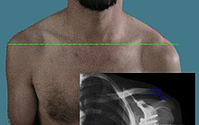
The cause is usually indirect trauma, such as a fall on the arm. The most common is shoulder dislocation , which accounts for more than 50% of all traumatic dislocations, followed by elbow dislocation . Dislocations of the knee or ankle are also common. Almost all joints can be affected (including jaw dislocation). Hyperextension injuries to the finger joints usually lead to dislocation, more often in handball and volleyball. The shoulder joint dislocation occurs most frequently when a bicycle falls (see picture). Rarely, direct traction can also trigger a dislocation, as in childhood dislocation of the radial head by pulling on the extended pronated arm ( pronatio dolorosa Chassaignac ).
During the examination, there is a relieving posture with loss of function and pain, occasionally swelling and a bruise. So-called “safe” signs of dislocation are a visible deformity, a recognizable empty joint socket and abnormal position of the joint head (often visible on the shoulder) and a resilient fixation. However, a dislocation can also be present with an apparently intact joint function.
X-rays in two planes provide evidence, although rare forms (such as the posterior shoulder dislocation) and child dislocations are difficult to detect. Computed tomography (CT), magnetic resonance imaging (MRT) or arthrography (special X-ray technology with the introduction of a contrast medium into the joint) can help . In children, dislocations can be easily visualized on ultrasound .
In the event of a traumatic dislocation, immediate reduction is required. This should always be done gently and not brusquely or with great force, as otherwise there is a risk of nerve and vascular damage and injuries to the joint. If relaxation is not possible, reduction is carried out under analgesic sedation or anesthesia . The repositioning must then be documented in the X-ray, followed by immobilization (on the shoulder e.g. in a Gilchrist bandage , on the ankle in the case of subtalar dislocation in a non-resilient short cast) and, if necessary, further examinations to exclude injuries to the bone parts, the joint capsule , the joint lip and the surrounding ligaments.
If a closed reduction is not possible or if there is a combination with a broken bone (dislocation fracture), the reduction is performed surgically with opening of the joint ( arthrotomy , so-called open or bloody reduction). Injured ligament structures (e.g. collateral ligaments) and accompanying fractures are usually treated surgically. The main complications are joint instabilities caused by tearing the joint capsule and the surrounding ligaments. This can result in further dislocations, including habitual dislocation (see below). A tear of the joint lip (on the shoulder: Bankart's lesion ) can lead to joint instability, often associated with a feeling of insecurity and fear of dislocating the joint again. Instability and repeated dislocations lead to premature osteoarthritis . Accompanying fractures can also occur, such as the impression fracture on the back of the humerus head ( Hill-Sachs lesion ) or dislocation fractures. Cartilage damage also plays a role in the long-term consequences. Violent repositioning can also damage blood vessels and nerves.
Central hip dislocation is a special form of traumatic hip dislocation . In the event of strong, axial force on the thigh, for example in car accidents at high speed and falls from great heights, the femoral head is driven through the shattered socket into the small pelvis. As with dislocation fractures, surgical treatment is necessary.
Habitual dislocation
Usually triggered by a traumatic initial dislocation, if the instability remains with less force and finally without any further accident mechanism, repeated dislocations, a so-called habitual dislocation, occur. It is most common after a shoulder dislocation and after a dislocation of the kneecap . Occasionally, the joint can be dislocated on request and independently reduced (set in) (so-called voluntary dislocation).
Congenital dislocation
The dislocation is already present at birth or develops from a congenital joint dysplasia . Hip dysplasia is most common in around 1–2% of all newborns and congenital hip dislocation in around 0.1% of all newborns. Congenital knee joint dislocation or congenital radial head dislocation are much less common . All joints can be affected, but this is very rare, but it can happen. a. in the context of Larsen's syndrome with multiple dislocated joints.
Chronic dislocation
Chronic diseases or malpositions lead to increasing destruction of the joints, which gradually leads to complete dislocation via subluxation (destructive dislocation). This is no more painful than the underlying disease or deformity. A repositioning on its own is usually not possible and makes no sense, since a new dislocation occurs immediately if there is a lack of stability. All joints can be affected. Typical examples are:
- Foot malposition with hallux valgus and contractures of the little toes ( hammer toe , claw toe )
- Destruction of the joint and ligament apparatus caused by a joint infection (septic arthritis )
- Rheumatism induced arthritis with destruction of the sidebands of the holding apparatus and the joint capsule (typically in the hands and feet)
- With flaccid and spastic paralysis, gradually increasing misalignment up to dislocation; with spasticity of the adductor muscles, often progressive hip subluxation
- As a result of a tumor near the joint
- In osteonecrosis with subsequent deformation of the adjacent joint, v. a. for femoral head necrosis . Usually only subluxation occurs. The more serious problem is usually osteoarthritis .
- Legacy traumatic, unreduced dislocation (most common radial head dislocation in children)
Lens dislocation
The Linsenluxation is complete (ectopia of the lens; ektopos = shifted) or partially (Linsensubluxation) displacement of the lens (for example, in the front. Oculi ). It can be congenital (e.g. in Marfan syndrome ) or acquired through an accident.
Tooth dislocations
In dentistry , a dislocation is a trauma-related abnormal change in position of a tooth ( total dislocation , subluxation , lateral dislocation ...).
The movements that the dentist uses to remove a tooth are also known as "dislocation movements".
swell
- KL Krämer, M. Stock. M. Winter: Clinical Guide Orthopedics . 3. Edition. Gustav-Fischer-Verlag, Ulm 1997.
- AM Debrunner: Orthopedics - Orthopedic Surgery . 3. Edition. Hans Huber Publishing House, Bern 1994.
See also
- Patellar luxation
- Radial head subluxation
- Shoulder dislocation
- Lens dislocation
- Hip dysplasia
- Ehlers-Danlos Syndrome
Individual evidence
- ↑ auskugeln in Duden (accessed April 25, 2018)
- ↑ Joachim Grifka, Jürgen Krämer: Orthopädie Unfallchirurgie Springer-Verlag, 2013, ISBN 978-3-642-28875-3 . P. 19 .
- ↑ Treatment and outcome of subtalar dislocations, dissertation, University of Würzburg, Sebastian Kiesel, July 2015, p. 30 ( PDF ).
- ↑ The subtalar dislocation - a dramatic event with dramatic consequences? In: German Congress for Orthopedics and Trauma Surgery. October 2, 2006, accessed January 10, 2018 .


