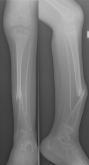
Congenital tibial osteoarthritis
| Classification according to ICD-10 | |
|---|---|
| Q74.9 | Unspecified congenital malformation of the extremity (s) Including: congenital anomaly of the extremity (s) onA |
| M84.1 | Non-union of the fracture ends [pseudarthrosis] |
| ICD-10 online (WHO version 2019) | |
A congenital or infantile tibial osteoarthritis ( Latin: Crus varum congenitum , English Congenital pseudarthrosis of the tibia ) is a rare special form of pseudarthrosis on the lower leg, which affects both the tibia and the fibula as well as the accompanying soft tissues and often in patients with Neurofibromatosis occurs.
Basically, it is an increasing curvature of the lower leg bones with infractions or fractures that do not heal, but rather show a tendency to thinning of the compacta and bone regression. The middle and / or distal third of the tibia is usually affected. Contrary to the usual name, pseudarthrosis is not innate, only the tendency to it.
The disease is a congenital malformation of the lower leg and often occurs together with hypoplasia of the tibia.
Epidemiology
The disease occurs in around 1: 200,000 births.
causes
A circumscribed disturbance of bone formation is assumed to be the cause. Histologically , neurofibromas or hamartoma-like tissue proliferation are found in the tissue of the pseudarthrosis .
Classification
Depending on the extent of the radiologically detectable change, a classification can be made, e.g. B. Crawford
- Type I crus antecurvatum, only antecurvation and cortical thickening
- Type II crus varum et antecurvatum, antecurvation + varus bending + sclerotherapy
- Type III cystic structural changes as in fibrous dysplasia , fractures in the first year of life
- Type IV dysplastic, bone thinned, sclerosed, thinned.
The last two forms are often found in neurofibromatosis.
diagnosis
The main symptom is anterior curvature and shortening of the lower leg, the diagnosis is made by means of an X-ray . The disease becomes noticeable in the first few years of life
The magnetic resonance imaging is used for preoperative planning, showing the exact extent of the changes to be resected. Typically, the abnormal tissue portions on T2-weighted sequences are signal-intensive and absorb contrast media.
The tibia recurvata is to be demarcated with a significantly more favorable course.
treatment
Treatment depends on the type and whether pseudarthrosis has already formed.
In this way, orthoses can prevent fracturing.
Only resection of the pseudarthrosis with the entire changed tissue helps causally (see #Causes).
A vascular pedicled transfer of the fibula on the opposite side, external fixator treatment and others are possible.
Due to the rarity of the disease and the complexity of the treatment, it should be done at a specialized center.
Individual evidence
- ↑ a b c J. P. Kuhn, TL Slovis, JO Haller (Ed.): Caffey's Pediatric Diagnostic Imaging. Volume 2, 10th edition. Mosby 2004, ISBN 0-323-01109-8 , p. 2111.
- ↑ pseudarthrose.ch
- ↑ a b c d e F. Hefti: Pediatric orthopedics in practice . Springer 1998, ISBN 3-540-61480-X .
- ↑ AHCrawford: Neurofibromatosis in children. In: Acta Orthopedica Scandinavica. Supplement 1986, (218), pp. 1-60.
- ↑ a b c H. Shah, M. Rousset, F. Canaves: Congenital pseudarthrosis of the tibia: Management and complications. In: Indian Journal of Orthopedics. 46, 2012, pp. 616-626. (online at: ijoonline.com )
- ↑ BM O'Brien, GJ Gumley, BJ Dooley, JJ Pribaz: Folded free vascularized fibula transfer. In: Plastic and reconstructive surgery. Volume 82, Number 2, August 1988, pp. 311-318, ISSN 0032-1052 . PMID 3399561 .
Web links
- Pseudarthrosis.ch
- H. Shah, M. Rousset, F. Canavese: Congenital pseudarthrosis of the tibia: Management and complications. In: Indian journal of orthopedics. Volume 46, number 6, November 2012, pp. 616-626, doi : 10.4103 / 0019-5413.104184 , PMID 23325962 , PMC 3543877 (free full text).
swell
- F. Hefti: Children's orthopedics in practice. Springer 1998, ISBN 3-540-61480-X .
- F. Hefti, G. Bollini, P. Dungl et al .: Congenital pseudarthrosis of the tibia: History, etiology, classification, and epidemiologic data. In: Journal of Pediatric Orthopedics. (B) 9, 2000, pp. 11-15.
