Magnetic resonance angiography

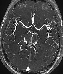
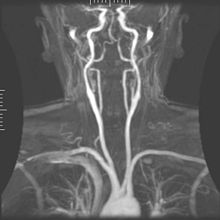
The magnetic resonance angiography (MR angiography, MRA) is an imaging method for the diagnostic visualization of blood vessels (arteries and veins) with the methods of magnetic resonance imaging (MRI). For this purpose, different techniques can be used, some of which are completely non-invasive (that is, they do not require surgery or injections) or are based on the administration of MRI contrast media . In contrast to conventional angiographyInstead of two-dimensional projection images, MRA generally records three-dimensional data sets that enable the vessels to be assessed from all directions. Another difference to conventional angiography is that MRA does not require a catheter to be inserted into the blood vessel system. In many areas, MRA is a competing procedure with digital subtraction angiography , CT angiography and sonography .
Indications
Typical indications for an MRA examination are, for example, suspected arterial stenoses ( atherosclerosis ) , vascular occlusions ( embolisms ) , venous thromboses , vascular sacs ( aneurysms ) , vascular malformations and other vascular diseases as well as the examination of the vascular conditions of tumors .
MRA techniques
There are many different techniques for using magnetic resonance imaging to visualize blood vessels. The main techniques are listed below:
Time-of-flight MRA
The time-of-flight MRA (TOF-MRA) takes advantage of the fact that freshly flowing blood has a higher magnetization in the examination volume than the stationary tissue, the magnetization of which is reduced ( saturated ) by the RF pulses of the MRT pulse sequence . The blood vessels with freshly flowing blood are therefore displayed with a high level of signals. Fast 2D or 3D gradient echo techniques ( FLASH ) are usually used for the TOF-MRA ; no contrast agent is required.
However, only vessels containing freshly flowing blood are shown as signal-rich. This can lead to artifacts if, for example, vessels run over long distances within the examination volume; in this case the magnetization of the blood will also become saturated over time. For this reason, the position of the examination volume cannot be chosen arbitrarily, but must generally be perpendicular to the prevailing blood flow direction.
The name Time-of-Flight-MRA refers to the fact that the magnetization of the blood took place outside the examination volume and a certain running time is required before the blood with higher magnetization reaches the area shown. German translations such as flight time or transit time MRA are rarely found.
Phase contrast MRA
The movement of the flowing blood can be represented as a phase difference in the complex image data using suitable examination techniques . Due to these phase differences, the flowing blood differs from the surrounding stationary tissue and can therefore be displayed with a rich signal. The phase contrast MRA (PC-MRA from English phase-contrast MRA , rarely also PK-MRA) is usually based on fast gradient echo techniques ( FLASH ) with additional flow-encoding gradient pulses and does not require a contrast agent.
In contrast to the time-of-flight MRA, the position of the examination volume is much more flexible with the phase contrast MRA. On the other hand, the recording times are much longer than with the time-of-flight MRA.
Contrast Enhanced MRA
By injecting a T1-shortening (mostly gadolinium-based ) contrast agent , the blood is displayed with a high level of signal on T1-weighted MRI images. MRT recordings while the contrast medium is flowing through an organ or an anatomical region can therefore be used to depict the blood vessels. The contrast -enhanced MRA (CE-MRA from English contrast-enhanced MRA , occasionally also KM-MRA from contrast agent MRA) is based on fast, usually three-dimensional gradient echo techniques ( FLASH ).
Compared to time-of-flight or phase contrast MRA, the recording time can be significantly shortened with contrast-enhanced MRA, so that recordings with held breath are just as possible as dynamic MRA recordings that can display the blood flow in a time-resolved manner (with time resolutions down to at 1 second per 3D data set).
More techniques

Spin echo techniques, which depict fast-flowing blood with low signal levels , can be combined with ECG triggering in order to display the arteries in two consecutive recordings, one with low signal (recording during fast systolic blood flow) and one with high signal (recording during slow diastolic blood flow). In the difference between the two recordings, the arteries appear signal-rich, while other tissue is suppressed.
Other techniques such as steady state free precession sequences or arterial spin labeling methods can also be used to display the blood in the vessels as rich in signals.
In contrast to the high-signal representation of the vessels, which is usually sought, there is also the option of displaying the vessels with low signal (dark) in the surrounding lighter tissue. This is used, for example, to display veins (MR phlebography ) with T2 * and susceptibility-weighted pulse sequences.
Post-processing and presentation of the MRA data
For assessment and diagnosis, the three-dimensionally recorded data records must be displayed as two-dimensional images (on the screen). There are various post-processing techniques for this: The data can be viewed in layers according to the original exposure orientation, they can be reconstructed as multiplanar reconstructions (MPR) in any planes (e.g. at right angles to the vessel or with the vessel in the image plane) , maximum intensity projections (MIP, maximum intensity projection ) can be calculated from different angles or the data can be displayed as virtual three-dimensional objects in space.
For all advanced display techniques, it is advantageous to record data that is as isotropic as possible , in which the spatial resolution is roughly the same in all three spatial directions. Typical spatial resolutions with modern techniques (depending on the size of the vessels of interest and the receiving volume) are between 1.5 × 1.5 × 1.5 mm³ and 0.5 × 0.5 × 0.5 mm³.
Individual evidence
- ↑ Konrad Meyne: Handbook of arterial occlusive diseases . Schlütersche, Hannover 2003, ISBN 3-87706-694-1 , p. 77 . ( limited preview in Google Book search)
- ↑ Eike Nagel u. a. (Ed.): Cardiovascular magnetic resonance tomography: understanding of methods and practical application . Steinkopff-Verlag, Darmstadt 2002, ISBN 3-7985-1285-X , p. 107 . ( limited preview in Google Book search)
- ^ Mathias Goyen (ed.): MR angiography with Vasovist . ABW Wissenschaftsverlag, Berlin 2007, ISBN 3-936072-52-3 . ( limited preview in Google Book search)
- ↑ Harald H. Quick: Magnetic resonance angiography: Basics and practice for MTRA . ABW Wissenschaftsverlag, Berlin 2007, ISBN 3-936072-46-9 , p. 3-67 . ( limited preview in Google Book search)
