Microphotography
As photomicrography an area which is Photography designated in which the images of objects on a sensor are much larger than the objects themselves. This occurs imaging scales are larger than that of macro photography .
A particular difficulty with microphotography is the very shallow depth of field . In the case of digital recordings of still objects, it is possible to create an image with increased depth of field by stacking several recordings with different focus planes (“ focus stacking ”). There is special software for this, such as the open source program CombineZP .
Microphotography is used professionally for documentation in the areas of biology (especially histology ), mineralogy and material testing.
Microphotography using a microscope
The most common microphotography is done with the help of a camera attached to a microscope.
Microscopes with a phototube
For this purpose, research microscopes usually have a phototube to which the camera housing is connected without additional lenses. In this case, the intermediate image completely illuminates the film or the sensor surface ( CCD / CMOS ) of the camera, the ocular optics are not involved in the recording. This arrangement offers numerous advantages:
- The quality of the recording is not impaired by additional optical elements,
- the camera is mechanically rigidly connected to the microscope,
- the eyepiece optics remain free for the selection of the image section,
- When using bayonet locks , the camera housing can be changed in a few seconds,
- Different recording formats can be used by adapting them using intermediate rings.
Most manufacturers of research microscopes also offer camera housings that are specially tailored to their own makes and thus enable better control of the exposure. Further developments in microphotography consist in the transfer of the digitized recordings to a workstation for software-controlled, automated image evaluation - not only of individual images, but also entire series, such as histological sections .
Microscopes without a phototube
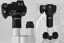
With microscopes without a photo tube, however, it is quite possible to hold a camera (even without further adjustments) directly to the eyepiece and release it with a steady hand. The alignment of the optical axis of the camera objective with that of the microscope eyepiece is particularly important . Not every camera is suitable for this, but it is possible with many inexpensive models. Due to a lack of optical adjustment, usually only a smaller image with vignetted (shadowed) corners and edges can be obtained.
A slightly better, but also more expensive solution is to use a tube adapter with which the camera is connected to the tube instead of the eyepiece . The adapter takes over the mechanical and optical adjustment between microscope and camera. This avoids blurring and vignetting of the image, the resulting photo quality is much higher.
Electron microscopes
While scanning electron microscopes can not record images directly, this is possible with transmission electron microscopes . Photo plates , sheet film or 35 mm film are traditionally used. Modern devices are equipped with CCD cameras , which can record the images directly in the microscope via a scintillator . Magnifications of over a million can be achieved with resolutions of up to 0.05 nm.
Web links
- Anja Hartmann: www.mikrofoto.de - Microscopy & Microphotography
- Eckart Hillenkamp: Use of the Nikon Coolpix 990 digital camera in microscopy at www.mikoskopieren.de
- Frank Fox: Professional microphotography and macro photography , many sample images of small organisms, plant cuttings and crystals.
- Digital cameras suitable for microscope photography , overview at MICRO TECH LAB.
- Photography with microscopes - adapter and repro column


