Osteoid osteoma
| Classification according to ICD-10 | |
|---|---|
| D16 | Benign neoplasm of bone and articular cartilage |
| ICD-10 online (WHO version 2019) | |
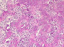
Osteoid osteomas are small and painful, but benign, bone tumors with self-limiting growth in size. Henry Lewis Jaffe identified the entity .
pathology
They consist of a heavily vascularized "core" ( nidus ), which is surrounded by a rounded or spindle-shaped zone, reactively growing, compacted (sclerotic) mature bone tissue. Preferably, osteoid osteomas are located in the upper and lower leg bones. If the nidus exceeds 1.5 cm, the change is called an osteoblastoma .
About 14 percent of all bone tumors are osteoid osteomas. The tumor usually occurs between the ages of 10 and 20 and is three times more common in males. Occurrence before the age of 10 is possible, very rarely after the age of 30. Osteoid osteomas show symptoms in the form of local pain, which tend to occur at night.
The diagnosis is made using imaging methods: X-rays , bone scintigraphy , CT and MRT (preferably as a dynamic examination with evidence of arterial contrast medium enrichment in the well-perfused nidus).
Differential diagnosis
therapy
Therapy consists of surgical removal by means of open resection or percutaneous punch biopsy or, preferably, CT-controlled radio frequency therapy . Alternatively, if an osteoid osteoma is present, symptomatic treatment with aspirin or other non-steroidal anti-inflammatory drugs can be used, since the disease is usually observed to heal within a period of about 2 to 7 years.
In June 2008, the first MRT-guided laser resection of an osteoid osteoma of the fibula was performed in the open high-field MRI of the Charité in Berlin-Mitte.
literature
- Jürgen Freyschmidt , Helmut Ostertag and Gernot Jundt: Bone tumors with jaw tumors. Clinic-Radiology-Pathology , 3rd edition. Springer, Berlin 2010. ISBN 978-3-540-75152-6 .
Web links
Individual evidence
- ^ E. Levine, JR Neff: Dynamic computed tomography scanning of benign bone lesions: preliminary results. Skeletal Radiology 9 (1983), pp. 238-245, ISSN 0364-2348 . PMID 6867773 .
- ^ PT Liu, FS Chivers, CC Roberts, CJ Schultz, CP Beauchamp: Imaging of osteoid osteoma with dynamic gadolinium-enhanced MR imaging. Radiology 227 (2003), pp. 691-700, ISSN 0033-8419 . doi : 10.1148 / radiol.2273020111 . PMID 12773675 .
- ↑ DI Rosenthal, FJ Hornicek, M. Torriani, MC Gebhardt, HJ Mankin: osteoid osteoma: percutaneous treatment with radiofrequency energy. In: Radiology 229 (2003), pp. 171-175, ISSN 0033-8419 . doi : 10.1148 / radiol.2291021053 . PMID 12944597 .
- ↑ K. Woertler, T. Vestring, F. Boettner, W. Winkelmann, W. Heindel, N. Lindner: Osteoid osteoma: CT-guided percutaneous radiofrequency ablation and follow-up in 47 patients. In: Journal of Vascular and Interventional Radiology 12 (2001), pp. 717-722, ISSN 1051-0443 . PMID 11389223 .
- ↑ T. Goto, Y. Shinoda, T. Okuma, K. Ogura, Y. Tsuda, K. Yamakawa, T. Hozumi: Administration of nonsteroidal anti-inflammatory drugs accelerates spontaneous healing of osteoid osteoma. In: Archives of Orthopedic and Trauma Surgery (2010), ISSN 1434-3916 . doi : 10.1007 / s00402-010-1179-z . PMID 20737157 .

