Pseudopod
False pods or pseudopodia (sing. Pseudopodium) are plasma protuberances of eukaryotic cells. In protists , they are morphologically very diverse and perform numerous functions, particularly in terms of movement and metabolism. They are also of great importance in cell migration in tissue animals .
Shapes of pseudopods
Basically five types of pseudopodia can be distinguished: lobopodia, filopodia, lamellipodia, reticulopodia and axopodia.
Lobopodia

Lobopodia are found particularly in Amoebozoa . They taper to a point or are tubular, tongue or finger-shaped and can change their shape very quickly. Inside, the plasma flows very quickly. They are formed either individually (monopodial types) or in larger numbers (polypodial types). Lobopodia are particularly useful for movement.
Filopodia
Filopodia can be found u. a. in radiation animals ( Radiolaria ) and are thread-like protuberances of the cell , usually straight, occasionally also curved, rarely weakly branched. There is no microtubule axoneme. In macrophages , filopodia act as tentacles that pull bound objects to the cell in order to phagocytose them .
Lamellipodia
Lamellipodia (occasional spelling variant: lamellopodia) are very flat and broad-based cell processes that are z. B. found in some Aconchulina .
Reticulopodia

Reticulopodia are special forms of pseudopodia, which can only be found in foraminifera as single-cell organisms with a predominantly stationary way of life.
The pseudopods branch out and can also merge again. They form extensive networks (pseudopodial networks) that serve to catch prey, to move around, to transport organelles within the cell and to anchor them underground. Reticulopodia always show granule flow and internal microtubules .
Axopodia
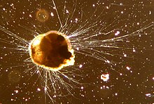
Axopodia, sometimes also called actinopodia, are found among other things in sun animals and radiation animals and are particularly long and straight cell processes with an axoneme made up of microtubules. In addition to catching prey, they serve to increase the water resistance, to slow down sinking and for locomotion.
Cellular processes in the formation of pseudopodia
The cellular processes should be illustrated using the example of amoebas: amoebas have an external, hyaline ectoplasm , which is in the gel state, and an internal granular endoplasm , which is in the sol state. In the boundary layer between the endoplasm and the ectoplasm there are actin and myosin filaments , the sliding and sticking of which leads to a stiffness of the affected area, similar to the contraction and relaxation of muscle cells . An actin binding protein (ABP) links actin to form a gel-like network. If the calcium content increases , gelsolin is released, which breaks down the actin filaments. As a result, the ectoplasm in the area of action of the gelsolin changes into the sol state and thus disappears (change in physical state). The actin-myosin interaction is resolved at the point. At the opposite end, the physiological end ( uroid ) of the cell, this interaction persists and contracts. As a result, the endoplasm is displaced and flows forward, where there is no ectoplasm, i.e. no gel-like covering. The endoplasm can pass through the actin-myosin network in this area, but not the granular inclusions and cell organelles . This plasma flow leads to the drawing in of pseudopodia on the uroid. This leads to the formation of folds in the cell membrane and the formation of new pseudopodia at the front end due to the cell membrane bulging. The pseudopodia only disappear again when the ABP rebuilds the actin filaments.
literature
- PK Mattila, P. Lappalainen: Filopodia: molecular architecture and cellular functions. In: Nature reviews. Molecular cell biology. Volume 9, Number 6, June 2008, pp. 446-454, ISSN 1471-0080 . doi : 10.1038 / nrm2406 . PMID 18464790 . (Review).
Individual evidence
- ↑ a b c d e Klaus Hausmann, Norbert Hülsmann, Renate Radek: Protistology , 3rd edition, Schweizerbart, 2003, p. 23, ISBN 3-510-65208-8 .
- ↑ a b c d e Rudolf Röttger: Dictionary of Protozoology In: Protozoological Monographs, Vol. 2, 2001, ISBN 3-8265-8599-2 .
- ↑ Klaus Hausmann, Norbert Hülsmann, Renate Radek: Protistology , 3rd edition, Schweizerbart, 2003, p. 146, ISBN 3-510-65208-8 .
- ↑ Klaus Hausmann, Norbert Hülsmann, Renate Radek: Protistology , 3rd edition, Schweizerbart, 2003, p. 213, ISBN 3-510-65208-8 .
- ↑ Kress H, Stelzer EH, Holzer D, Buss F, Griffiths G, Rohrbach A: Filopodia act as phagocytic tentacles and pull with discrete steps and a load-dependent velocity . In: Proc. Natl. Acad. Sci. USA . 104, No. 28, 2007, pp. 11633-11638. doi : 10.1073 / pnas.0702449104 . PMID 17620618 . PMC 1913848 (free full text).
- ↑ a b Keyword “Reticulopodia.” In: Herder-Lexikon der Biologie. Spectrum Akademischer Verlag GmbH, Heidelberg 2003. ISBN 3-8274-0354-5
- ↑ Volker Storch, Ulrich Welsch: Kükenthal zoological internship , ISBN 3-8274-1643-4 , 2005
