
Frontal sinus


The frontal sinus ( sinus frontalis ) is one of the paranasal sinuses ( sinus paranasales ). It represents a cavity in the frontal bone ( os frontale ) lined with mucous membrane , which is connected to the middle nasal passage ("sinus passage") of the nasal cavity . In addition, there is a connection to the ethmoid cells , which is particularly spacious in horses , so that both sinuses in this species are also summarized as the conchofrontal sinus . The frontal sinuses on both sides are separated from each other by a thin septum ( septum sinuum frontalium ).
Species specifics
The size and shape of the frontal sinus vary greatly. This applies not only to the comparison between different species , but also between different individuals in humans. There are four basic shapes described for humans: bean, leaf, mitral and pyramid shape. The size of the human frontal sinus varies between 0.05 and 7.8 cm 3 , and it is not uncommon for the frontal sinus not to develop at all ( aplasia ) or only slightly ( hypoplasia ). The bony roof of the eye socket can also be pneumatized to a variable extent by the frontal sinus.
In cattle and pigs , the frontal sinus is particularly large and extends to the occiput ( Os occipitale ). In horn-bearing ruminants , the frontal sinus has a branch into the horny process ( processus cornualis ) of the skull. Therefore, when the horn is broken off or when adult ruminants are dehorned, the frontal sinus is also opened.
development
In newborns, the frontal sinus cannot be separated from the ethmoid cells. Pneumatization of the frontal bone does not begin in humans until the age of 2, and in seven to eight year olds it reaches the upper edge of the eye socket.
Diseases
Like the other paranasal sinuses, the frontal sinus can also be affected in diseases of the nose. Inflammation of the frontal sinus is called sinusitis , and a collection of pus is called frontal sinus empyema .
In the case of illnesses or accumulation of secretions, a frontal sinus can be rinsed. In chronic cases, radical frontal sinus surgery, i.e. H. surgical removal of the diseased frontal sinus mucosa may be indicated.
In humans, osteomas are occasionally found in the frontal sinus.
In a fracture of the frontal sinus at a participation of the rear wall is the risk of infection of the intracranial ( meningitis or encephalitis ) and / or the development of a CSF fistula added.

Impression fracture of the frontal sinus in computed tomography (arrow). The rear wall of the frontal sinus leading to the skull is not affected here.
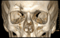
Impression fracture of the frontal sinus in computed tomography: 3D reconstruction using volume rendering technology .

Some sinuses, bony structures


literature
- Franz-Viktor Salomon: Bony skeleton. In: Franz-Viktor Salomon, Hans Geyer, Uwe Gille (Ed.): Anatomy for veterinary medicine. Enke, Stuttgart 2004, ISBN 3-8304-1007-7 , pp. 37-110.
- Theodor H. Schiebler (Ed.): Anatomie. Histology, history of development, macroscopic and microscopic anatomy, topography. Taking into account the item catalog. 9th, completely revised edition. Springer, Berlin et al. 2005, ISBN 3-540-21966-8 .
Individual evidence
- ^ A b Anton Waldeyer : Human anatomy. 17th, completely revised edition. Walter de Gruyter, Berlin et al. 2003, ISBN 3-11-016561-9 , p. 324.
- ↑ H. Wechalekar: Extension of the frontal sinus into the roof of the optic canal: a cadaveric case report. In: Surgical and Radiologic Anatomy Volume 38, No. 5, 2016, pp. 609-613. doi: 10.1007 / s00276-015-1560-2 ; accessed on March 8, 2018