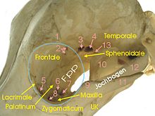
Orbit

yellow = frontal bone
green = tear bone
brown = ethmoid bone
blue = cheekbone
violet = upper jaw
light blue = palatine bone
red = sphenoid bone
Orbita (from Latin orbis 'circle'; plural orbitae ) denotes the bony eye socket , a deep pit on the skull ( cranium ) in which the eye and its appendage organs are located. The anatomical term was coined by Gerhard von Cremona in the twelfth century when he translated Avicenna's canon of medicine into Latin. In humans, the pit is about 4 to 5 cm deep.
Involved bones
The orbit is made up of seven bones:
- Frontal ( frontal )
- Lacrimal bone ( os lacrimale )
- Upper jaw ( maxilla )
- Zygomatic bone ( os zygomaticum )
- Ethmoid ( ethmoid )
- Palatine bone ( os palatinum ) and
- Sphenoid ( sphenoid ).
In most mammals, the orbit is bony all around. In predators and pigs , the lateral edge of the temporal fossa ( fossa temporalis ) is only closed by a connective tissue band ( ligamentum orbitale ). It runs between the zygomatic process of the frontal bone and the frontal process of the zygomatic bone (see picture Hund, No. 1 and No. 9).
The distance between the two eye sockets is called the interorbital space.
Openings inside
There are several openings on the inner walls for nerves and blood vessels as well as the tear duct to pass through.
- Ethmoid foramen (sometimes several openings): for the passage of the vessels and nerves of the same name
- Optic Canal : for the passage of the optic nerve ( N. opticus , II) and the ophthalmic artery
- Fissura orbitalis ( superior in humans): for the passage of the other cranial nerves for the eye muscles and the sensitive innervation of the globe ( oculomotor nerve (III), trochlear nerve (IV), ophthalmic nerve (V1), abducens nerve (VI))
- Fissura orbitalis inferior (human only): for the passage of the vena ophthalmica inferior, nervus zygomaticus and nervus infraorbitalis (in animals foramen rotundum or foramen orbitorotundum )
- Fossa sacci lacrimalis and foramen lacrimal : tears, through the hole it goes into the tear-nasal duct to the nasal cavity (see tear ducts )
- Maxillary foramen : to the infraorbital canal for the nerves and vessels of the same name , leads to the infraorbital foramen
See also
Web links
literature
- Franz-Viktor Salomon: Bony skeleton . In: Franz-Viktor Salomon, Hans Geyer, Uwe Gille (Ed.): Anatomy for veterinary medicine. Enke, Stuttgart et al. 2004, ISBN 3-8304-1007-7 , pp. 37-110.
Individual proof
- ↑ Luis-A Arráez-Aybar: Toledo School of Translators and their influence on anatomical terminology . In: Annals of anatomy-Anatomischer Anzeiger . 198, August. doi : 10.1016 / j.aanat.2014.12.003 .

