Trochlear nerve


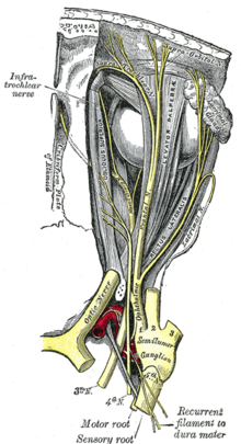
The paired trochlear nerve , also known as the fourth cranial nerve , N. IV , innervates the outer eye muscle Musculus obliquus superior (referred to in animals as the Musculus obliquus dorsalis ) and also carries afferent fibers from its proprioceptors ( deep sensitivity ) in its orbital section .
course
The cranial nerve nucleus of the trochlear nerve, the nucleus nervi trochlearis , lies in the midbrain and joins the main nucleus of the oculomotor nerve caudally . The fibers descend from the trochlear nucleus in the laterocaudal direction, pull around the central gray cave in a dorsal direction and cross in the trochlear decussatio to the opposite side, immediately before their brain exit. The trochlear nerve emerges as the only cranial nerve dorsally from the brain below the inferior (or caudal) colliculi of the quadrilateral plate and to the side of the rostral (or superior ) frenulum veli medullaris .
In the cisterna ambiens of the subarachnoid space, it winds laterally around the cerebral leg ( Pedunculus cerebri ) to the base of the brain stem and runs between the posterior cerebral artery and the superior cerebelli artery . At about the level of the Turkish saddle rest ( Dorsum sellae [turcicae] ) it penetrates the dura at the edge of the tentorium slit ( Incisura tentorii ) and runs in the side wall of the venous cavernous sinus . Here the trochlear nerve (N. IV) lies between the nervus oculomotorius (N. III) and the nervus ophthalmicus (N. V 1 ).
After a long intra- dural and the longest intra- cranial course of all cranial nerves, the thin N. IV leaves the cranial cavity in humans through the superior orbital fissure - in non-primates through the corresponding orbital fissure , in cloven-hoofed animals through the foramen orbitorotundum - and passes over the annulus tendineus communis in the eye socket to the only muscle innervated by it. The orbital course is also accompanied by afferent proprioceptive fibers, which - as with the other external eye muscles - reach the trigeminal ganglion via connections (anastomoses) with the ophthalmic nerve .
The tendon of the superior oblique muscle is deflected in the direction of its pull by a roll cartilage . This roll cartilage ( trochlea ) gave the nerve its name.
affection
Damage to the trochlear nerve leads to paralysis of the equilateral (ipsilateral) superior oblique muscle ( trochlear palsy ). The loss of function manifests itself in restricted eye movement and a squint , which can be different depending on the type (unilateral or bilateral paralysis) and direction of view and is associated with corresponding double vision ( diplopia ).
In this case, the affected eye deviates upwards ( hypertropia ) and inwards ( esotropia ). There is also an outward curling around the sagittal axis ( excyclotropy ). Double images occur depending on the direction of gaze, especially vertical ones when looking down, rotational ones increasingly when looking towards the affected side. The patient therefore often tilts his head to the side of the healthy eye to compensate ( Bielschowsky phenomenon ). This leads to a so-called ocular torticollis ( head posture or torticollis ocularis ).
In the case of isolated one-sided damage to the cranial nerve nucleus ( nucleus nervi trochlearis ), it should be noted that, due to the complete crossing of the nerve tracts, only the opposing (contralateral) muscle is affected.
A compression of the nerve on its intradural route is also possible through adjacent arterial vessels such as the posterior cerebral artery ( A. cerebri posterior ) or the upper cerebellar artery ( A. cerebelli superior ), which can lead to the rare picture of superior oblique myocymia ( nerve compression syndrome) ).
The differential diagnosis of congenital trochlear palsy also includes developmental anomalies in which the superior oblique eye muscle is malformed or its muscle mass is atrophied as part of a congenital cranial malnervation syndrome in the absence of a trochlear nerve. The forms of congenital strabismus associated with these rare clinical pictures are clinically described as decompensated strabismus sursoadductorius .
literature
- Martin Trepel: Neuroanatomy. Structure and function. 3rd, revised edition. Urban & Fischer, Munich et al. 2004, ISBN 3-437-41297-3 .
- Franz-Viktor Salomon: nervous system, systema nervosum . In: Franz-Viktor Salomon, Hans Geyer, Uwe Gille (Ed.): Anatomy for veterinary medicine. Enke, Stuttgart 2005, ISBN 3-8304-1007-7 , pp. 464-577.
- Herbert Kaufmann : Strabismus. 4th fundamentally revised and expanded edition, with Heimo Steffen., Georg Thieme Verlag, Stuttgart, New York 2012, ISBN 3-13-129724-7
Individual evidence
- ↑ Benninghoff: Macroscopic and microscopic anatomy of humans. Volume 3, Urban and Schwarzenberg, Munich 1985, ISBN 3-541-00264-6 , p. 440.
- ↑ SeokHoon Kang, Ji-Soo Kim, Jeong-Min Hwang, Byung Se Choi, Jae-Hyoung Kim: Mystery Case: Superior oblique myokymia due to vascular compression of the trochlear nerve. In: Neurology. Volume 80, No. 13, March 2013, doi: 10.1212 / WNL.0b013e318289706f .
- ↑ K. Scharwey, T. Krzizok, M. Samii, S. Rosahl, H. Kaufmann: Remission of superior oblique myokymia after microvascular decompression. In: Ophthalmologica. Volume 214, No. 6, 2000, pp. 426-428, PMID 11054004 , doi: 10.1159 / 000027537 .
