Tonometry
With tonometry (of TONOS, τόνος , Greek "pressure, voltage," and metron, μἐτρον , Greek "dimension") is the measurement of the intraocular pressure referred whose increase is usually one of the most important over the normal value addition, but not the only , a risk factor for glaucoma ( glaucoma group). It is measured in mmHg and is between 10 and 21 mmHg in a healthy adult. Procedures in which there is direct contact between the eye and the measuring device, the tonometer , first require local anesthesia of the cornea with drops so that the measurement is painless. As the intraocular pressure at different times may have different heights, this is with a corresponding indication measured repeatedly throughout the day and night, and a so-called days pressure profile summarized. The evaluation of the measurement results is reserved for ophthalmologists. A glaucoma can be present even when the intraocular pressure within the g o.. Normal range, and an increased intraocular pressure outside the normal range justifies suspicion of glaucoma .
Measurement method
Applanation tonometry
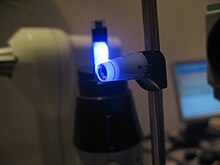
A common and precise method is applanation tonometry ( planus , Latin: "flat, even, flat") developed by the Austrian-Swiss ophthalmologist Hans Goldmann . A small measuring body is attached to a special ophthalmological examination device , the slit lamp . During the examination, the force is measured which is necessary to bring its flat front surface with a diameter of 3.06 mm (area = 7.35 mm²) into contact with the cornea and to flatten it. To visually check the contact between the cornea and the measuring body by the examiner, an aqueous solution of the fluorescein dye is instilled into the conjunctival sac. The force applied is generated by a spring balance that is coupled to a measuring drum. The pressure can be read off directly from this. There are also applanation tonometers that are hand-held in order to be able to carry out an inspection on, for example, recumbent persons.
Various studies refer to a correlation between corneal thickness and the pressure values determined in applanation tonometry . For people with a very thick cornea, the measurement result could then be incorrectly too high, for people with a very thin cornea it could be too low. In particular, after refractive operations in which the cornea was “thinned”, such measurement errors should occur. It is therefore recommended, especially in the case of corresponding patient groups, to determine the corneal thickness before measuring the intraocular pressure. This is usually done with a so-called pachymeter . The correct value can then be determined using a conversion factor using a correction table. However, other studies come to the conclusion that applanatory measured pressure values are not an artifact in patients with a thick cornea , and recommend, in case of doubt, a pressure value determination using an intraocular pressure probe .
However, other corneal properties such as rigidity and viscoelasticity also have an influence on the accuracy of the measurement of intraocular pressure. If necessary, other corneal-independent measurements are recommended.
Impression tonometry
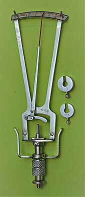
An older method of intraocular pressure measurement is impression tonometry ( impressio , Latin: "penetration, impression"). The Schiötz tonometer usually used for this , named after its developer, the Norwegian ophthalmologist Hjalmar August Schiøtz (1850–1927), is a simple device that is placed by hand on the anesthetized cornea of the patient lying on their back. It is shown how deep a metal pin with a defined weight indented the cornea. Scale values, weights used and the results obtained are listed in a calibration table. One problem with impression tonometry is that the instruments used are only calibrated for eyes with an average ability to stretch in the sclera ( rigidity ). In the case of abnormal rigidity (for example in the case of myopia ) the calibration table shows incorrect values for the intraocular pressure. For this reason, applanation tonometry is the method of choice.
Non-Contact Tonometry (NCT)
A procedure in which there is no contact between the eye and the measuring instrument is non-contact tonometry (NCT). The intraocular pressure is measured by means of an impulse with briefly increased air pressure . In terms of the examination process, this method is more pleasant for the test person, but because of the lower measurement accuracy, which is not systemically caused by the apparatus , it is not entirely meaningful.
While impression and applanation tonometric examinations are exclusively an ophthalmological activity, inspections and screenings of intraocular pressure by means of NCT are increasingly being used by opticians .
Transpalpebral Scleral Tonometry
Transpalpebral Scleral Tonometry refers to the method of measuring intraocular pressure through the eyelid. It avoids direct contact with the cornea and works according to the recoil principle, but through the eyelid (transpalpebral). The user places the device on the upper eyelid and initiates the measurement. The function of the sensor is performed by a freely movable rod that, when lowered, interacts with the elastic surface of the eye in the area of the sclera . The patient can sit or lie down, but must look up at a 45 ° angle. Local anesthesia is not required when using this method. With this method, the results of the intraocular pressure measurement are independent of the biomechanical condition of the cornea and should not be corrected with a pachymeter.
Dynamic Contour Tonometry
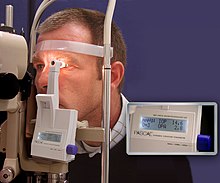
A new method is the so-called Dynamic Contour Tonometry (DCT). The dynamic measuring principle differs fundamentally from static applanation tonometry because it does not flatten the cornea, but rather the corneal measuring head brings the cornea into its natural, tension-free state. The curvature of the cornea under the measuring head is only slightly reduced (flatter). The pressure between the measuring head and the cornea then corresponds to the intraocular pressure. A pressure sensor built into the tonometer head can record the eye pressure directly and largely independently of corneal influences. The precision achieved makes it possible to display pulse curves of the intraocular pressure, which are triggered by the heartbeat, similar to an EKG .
The accuracy was confirmed in vivo in intracameral comparison measurements. It was shown that the DCT measurements correlate better than with an error of 1 mmHg with the actual intraocular pressure and that the corneal thickness has only a minimal influence on the measurement. The repeatability and reproducibility of the dynamic DCT measurements also turned out to be better than with the common static applanation tonometers or with the non-contact tonometers for single impulses.
After eye operations such as LASIK or crosslinking, which change the properties of the cornea, the results of DCT measurements are apparently less distorted than with applanation methods.
Eyemate
The process of continuous measurement over 24 hours and over long periods of time is still being researched, which is supposed to enable a microsensor called Eyemate , which received CE approval in 2017, and a number of at the University Eye Clinics in Aachen and Bochum selected patient has been successfully implanted. The intraocular pressure values are transmitted from the chip positioned in the anterior chamber to a reading device held by the patient at eye level and then telemetrically transmitted to the treating ophthalmologist. This method promises better control of the massive pressure fluctuations that frequently occur over the day and night.
Line of sight tonometry
A direction-dependent intraocular pressure measurement is carried out for the diagnostic assessment of the clinical picture of endocrine orbitopathy . The background to this investigation is that an inflammatory increase in the volume of the eye muscles caused by this disease can lead to compression of the eyeball when the direction of gaze is changed and the intraocular pressure can rise as a result. The same symptoms can occur with an orbital floor fracture .
Tonography
In contrast to the daily pressure curve, a measurement of the aqueous humor outflow carried out constantly over a period of a few minutes and the recording of a corresponding curve of the resulting pressure drop is called tonography . The procedure is considered a reliable method for the early detection of glaucoma simplex .
costs
The measurement of intraocular pressure is judged controversially as a precautionary measure : While the medical service of the umbrella association of health insurance funds assesses it as "tending to be negative", the director of the University Eye Clinic Mainz Norbert Pfeiffer believes "a general screening [is] absolutely sensible." scientifically oriented German Ophthalmological Society advocates the glaucoma early detection examination, in which the tonometry is included. The Robert Koch Institute wrote in 2017: ". An early diagnosis of glaucoma is important because apparent structural changes in the optic nerve head occur before the functional impairment" The costs are not covered by public health insurance in Germany, they that individual Represents health service (IGeL). However, if the measurement of intraocular pressure is medically necessary, for example after glaucoma has already been reliably diagnosed or in the case of long-term cortisone therapy, the costs are covered by the treatment flat rate. As a reason for the fact that the costs are not generally covered in the context of a glaucoma screening , the Federal Joint Committee states the "unknown reliability of the test in people without specific complaints or risk factors".
Web links
literature
- Theodor Axenfeld (founder), Hans Pau (ed.): Textbook and atlas of ophthalmology. With the collaboration of Rudolf Sachsenweger and others 12th, completely revised edition. Gustav Fischer, Stuttgart et al. 1980, ISBN 3-437-00255-4 .
Individual evidence
- ↑ Johannes Rückher: Tonometry : Measurement of the intraocular pressure. apotheken-umschau.de, December 4, 2014, accessed on March 5, 2016 .
- ^ Robert L. Stamper, Marc F. Lieberman, Michael V. Drake: Becker-Shaffer's Diagnosis and Therapy of the Glaucomas. 8th edition. Mosby, Elsevier, Edinburgh 2009, ISBN 978-0-323-02394-8 , pp. 47-50.
- ↑ Patient information from the Eye Clinic of the University Hospital Dresden
- ↑ Series of investigations by the University Eye Clinic Freiburg, connection between corneal thickness, applanation tonometry and directly determined intraocular pressure values
- ↑ 4th World Glaucoma Consensus Report on IOP
- ^ Peter WJ Dekker, Hartmut Kanngiesser, Yves C. Robert: The contact glass tonometer. In: Clinical monthly sheets for ophthalmology. Volume 208, No. 5, 1996, pp. 370-372, doi: 10.1055 / s-2008-1035243 .
- ↑ Hartmut E. Kanngiesser, Christoph Kniestedt, Yves C. Robert: Dynamic contour tonometry: presentation of a new tonometer. In: Journal of Glaucoma. Volume 14, No. 5, October 2005, pp. 344-350. PMID 16148581 .
- ↑ Andreas G. Boehm, Anja Weber, Lutz E. Pillunat, Rainer Koch, Eberhard Spoerl: Dynamic Contour Tonometry in Comparison to Intracameral IOP Measurements. In: Investigative Ophthalmology & Visual Science. Volume 49, No. 6, June 2008, pp. 2472-2477, doi: 10.1167 / iovs.07-1366 .
- ↑ Aachal Kotecha, Edward White, Patricio G. Schlott man, David F. Garway-Heath Intraocular pressure measurement precision with the Goldmann applanation, dynamic contour, and ocular response analyzer tonometer. In: Ophthalmology. Volume 117, No. 4, April 2010, pp. 730-737, doi: 10.1016 / j.ophtha.2009.09.020 .
- ↑ Antonios P. Aristeidou, Georgios Labiris, Andreas Katsanos, Michalis Fanariotis, Nikitas C. Foudoulakis, Vassilios P. Kozobolis: Comparison between Pascal dynamic contour tonometer and Goldmann applanation tonometer after different types of refractive surgery. In: Graefe's Archive for Clinical and Experimental Ophthalmology. Volume 249, No. 5, May 2011, pp. 767-773, doi: 10.1007 / s00417-010-1431-9 .
- ↑ MG Gkika, G. Labiris, VP Kozobolis: Tonometry in keratoconic eyes before and after riboflavin / UVA corneal collagen crosslinking using three different tonometer. In: European journal of ophthalmology. Volume 22, number 2, 2012 Mar-Apr, pp. 142–152, doi : 10.5301 / EJO.2011.8328 (currently unavailable) . PMID 21574162 .
- ↑ A. Koutsonas, P. Walter, G. Roessler, N. Plange: Long-term follow-up after implantation of a telemtric intraocular pressure sensor in patients with glaucoma: a safety report. In: Clin Exp Ophthalmol. 2017, published online on November 14th. DOI: 10.1111 / ceo.13100in
- ↑ Ronald D. Gerste: Glaucoma: Miniaturization in diagnostics and therapy. In: Deutsches Ärzteblatt. 113 (39), 2016, pp. A-1710 / B-1446 / C-1422.
- ↑ the manufacturer's website Implandata Ophthalmic Products GmbH
- ↑ University Hospital of the Ruhr-Universität Bochum - advanced training event for ophthalmologists on options for glaucoma therapy, 23 November 2016
- ↑ Herbert Kaufmann (Ed.): Strabismus. With the collaboration of Wilfried de Decker et al. 3rd, fundamentally revised and expanded edition. Georg Thieme, Stuttgart et al. 2003, ISBN 3-13-129723-9 , p. 430.
- ^ Theodor Axenfeld (founder), Hans Pau (ed.): Textbook and atlas of ophthalmology. With the collaboration of Rudolf Sachsenweger and others 12th, completely revised edition. Gustav Fischer, Stuttgart et al. 1980, ISBN 3-437-00255-4 , p. 446.
- ↑ IGeL-Monitor: Measurement of intraocular pressure for early detection of glaucoma , accessed on December 3, 2012.
- ↑ a b Jochen Niehaus, Regina Albers: IGeL: Health costs extra , focus.de/Focus No. 21, May 19, 2008, accessed on October 14, 2012.
- ↑ Statement of the German Ophthalmological Society on glaucoma screening. (PDF) August 2015, accessed on May 21, 2017 .
- ^ RKI: Federal Health Reporting. (PDF) In: GBE-themed booklet Blindness and Visual Impairment. Robert Koch Institute, 2017, accessed on May 21, 2017 .
- ↑ Individual health services (IGeL) - examination for the early detection of glaucoma. An information sheet from the statutory health insurance companies (PDF; 15 kB).
- ↑ Federal Joint Committee: Glaucoma screening does not make sense . Press release. April 5, 2005, accessed October 14, 2012.


