Chorion
The chorion (from ancient Greek χόριον, skin, leather, afterbirth '; also called serosa ) is the outer of the two fruit sheaths of the embryo or fetus of the "higher" land vertebrates (amniota). The space enclosed by the chorion is also known as the chorion or fruit cavity.
Who also "Chorion" said part of insect - cephalopods - and fish eggs is neither homologous nor similarly the Chorionic the amniotes because he no pericarp, but only an egg case is that the still in the ovary of the mother of the egg itself or by Follicular epithelial cells is deposited. Instead, the chorion of fish eggs is homologous to the zona pellucida of terrestrial vertebrate eggs .
Placental mammals
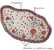
In Theria , the exchange of substances between mother and embryo or fetus ( uteroplacental circulation ) takes place, among other things, via the chorion. In the placental mammals it is chorion known and consists of the extraembryonic mesoderm and the trophoblast, the outer cell layer of the blastocyst (a very early but already poorly differentiated embryonic stage, which, inter alia, an embryonic and a adembryonalen pole has) which for this to Point in time has already differentiated into the outer syncytiotrophoblasts and the inner cytotrophoblasts. The chorion sends out villi in the direction of the endometrium , which after the implantation of a fertilized "egg cell" (more precisely: the blastocyst) is called Decidua graviditatis and surrounds the embryo in the form of the Decidua capsularis .
Villi formation begins in the blastocyst stage after the initial establishment of the uteroplacental circulation with the ingrowth of the primary villi from the cytotrophoblast into preformed, radial invaginations ( trabeculae ) of the syncytiotrophoblast. The trabeculae or primary villi protrude into a cavity of the syncytiotrophoblast ( lacunae ) into which the maternal blood capillaries open. From the inside, the extraembryonic mesoderm grows into the primary villi, which are called secondary villi from this stage on.
Although in this early phase of embryonic development the formation of lacunae, trabeculae and villi takes place over the entire chorion, it is particularly weak at the adembryonic pole, which points towards the uterine lumen . Later, villi formation is limited exclusively to the embryonic pole, while the villi regress in the remaining part of the chorion-decidua complex. That part of the chorion to which villi formation is then limited and where it continues is called chorion frondosum ('villi-rich chorion'), the part in which the villi recede and finally disappear is called chorion laeve ('smooth chorion') ) called. The chorion frondosum is the embryonic part of the placenta .
While the chorion differentiates itself into chorion frondosa and chorion laeve, capillary vessels form in the non-degenerating secondary villi through which embryonic, later fetal, blood flows. Later, these villi, now called tertiary villi, form numerous branches and the syncytiotrophoblast numerous microvilli , so that the increase in surface area between mother and fetus can be managed through the increased surface area . The space around the villi that is filled with maternal blood, which corresponds to the lacunae of early embryonic development, is called the intervillous space in the fully developed placenta .
See also
literature
- Hartmut Greven: Reproduction and Development. P. 167-182 in: Wilfried Westheide, Gunde Rieger (Ed.): Special Zoology. Part 2: vertebrates or skulls. 2nd edition, Spektrum Akademischer Verlag, Heidelberg 2010, ISBN 978-3-8274-2039-8
- KV Hinrichsen: Embryological basics. P. 30–61 in: Christof Sohn, Sevgi Tercanli, Wolfgang Holzgreve (ed.): Ultrasound in gynecology and obstetrics. 2nd, completely revised edition. Thieme, Stuttgart 2003, ISBN 3-13-101972-7 , p. 39 ff.
- Johannes W. Rohen, Elke Lütjen-Drecoll: Functional Embryology - The Development of the Functional Systems of the Human Organism. 3rd, revised and expanded edition. Schattauer, Stuttgart / New York 2006, ISBN 978-3-7945-2451-8
Web links
- Development of the Placental Villi - Human Embryology: Embryogenesis, Module 10: Fetal Membranes and Placenta. Joint website of the Universities of Friborg, Lausanne and Bern
- Placenta and Extraembryonic Membranes - University of Michigan Medical School website
Individual evidence
- ↑ Chris P. Raven: Oogenesis: The Storage of Developmental Information. Pergamon Press, 1961, p. 43 f.
- ^ Anne-Katrin Eggert, Josef K. Müller, Ernst Anton Wimmer, Dieter Zissler: Reproduction and Development. P. 363–459 in: Konrad Dettner, Werner Peters (Ed.): Textbook of Entomology. 2nd edition, Spektrum / Elsevier, Munich 2003, ISBN 3-8274-1102-5 , p. 369
- ↑ Kenji Murata, Fred S. Conte, Elizabeth McInnis, Tak Hou Fong, Gary N. Cherr: Identification of the Origin and Localization of Chorion (Egg Envelope) Proteins in an Ancient Fish, the White Sturgeon, Acipenser transmontanus. Biology of Reproduction. Vol. 90, No. 6, 2014, Item No. 132, doi: 10.1095 / biolreprod.113.116194 (Open Access).
- ↑ Birgit C. Hanusch: Anatomy. 1. General embryology. P. 152–168 in: Hamid Emminger (Ed.): Physikum EXAKT: the entire examination knowledge for the 1. ÄP. 4th, revised and updated edition. Thieme, Stuttgart 2005, ISBN 3-13-107034-X , p. 158 ff.
- ^ Franz Pera, Heinz-Peter Schmiedebach: Medical vocabulary. Compact terminology. 2nd Edition. De Gruyter, Berlin / New York 2010, ISBN 978-3-11-022694-2 , p. 38.


