Aortic aneurysm
| Classification according to ICD-10 | |
|---|---|
| I71 | Aortic aneurysm and dissection |
| I71.1 | Thoracic aortic aneurysm, ruptured |
| I71.2 | Thoracic aortic aneurysm with no indication of rupture |
| I71.3 | Abdominal aortic aneurysm, ruptured |
| I71.4 | Aneurysm of the abdominal aorta, with no indication of rupture |
| I71.5 | Aortic aneurysm, thoracoabdominal, ruptured |
| I71.6 | Aortic aneurysm, thoracoabdominal, no rupture indicated |
| ICD-10 online (WHO version 2019) | |

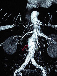
An aortic aneurysm is a bulging ( aneurysm ) of the main artery ( aorta ). One differentiates between aneurysms of the aorta at the level of the chest and abdominal (abdominal) variants. If the aneurysm is advanced, there is a risk of rupture with a high mortality rate.
Abdominal aortic aneurysm
An abdominal aortic aneurysm (BAA), abdominal aortic aneurysm (AAA) or aneurysm verum aortae abdominalis is considered to be an enlargement of the abdominal aorta below the exit of the renal arteries with an anterioposterior diameter of over 30 mm. Clinically, a distinction is made between asymptomatic, symptomatic and ruptured aneurysms. An asymptomatic (pain-free) aneurysm is an incidental finding. In the symptomatic aneurysm, the symptoms and in the ruptured the circulatory situation are in the foreground.
Symptoms
The symptoms of an abdominal aortic aneurysm are difficult to recognize and easy to confuse with other conditions such as acute heart attack . Diffuse abdominal and back pain, a poorly palpable, unevenly strong groin pulse, and dizziness can be symptoms. A pulsating tumor may be palpable as a sign of the bulge in the abdomen. The danger from the aneurysm comes from the possibility of rupture. The aorta located in the retroperitoneal space ruptures, but the bleeding is held back by the peritoneum and may progress unnoticed for several days. Acute annihilation pain in the abdominal area is described by those affected . In addition, symptoms of shock with a drop in blood pressure, subjective shortness of breath , fear of death and ischemic symptoms in the lower extremities can occur. These symptoms can be more or less pronounced and make it very difficult to distinguish them from a heart attack or arterial vascular occlusion in the preclinical setting. A symptom-free life with an aortic aneurysm is also possible, so many aneurysms are only discovered by chance during routine examinations.
In the laboratory examination of the blood values, a greatly increased D-dimer is found in an acute aortic aneurysm . Conversely, this can also be used as an exclusion criterion, since if the D-dimer is not at all or only slightly elevated, an aneurysm is most likely not present. This can be explained by the strong coagulation activity in the case of an acute aortic aneurysm, since there is a tear in the vascular wall that the body tries to close.
diagnosis
The diagnosis of AAA can be confirmed using various imaging methods .
- The ultrasound scan can find an AAA with a sensitivity and specificity of 90%. Due to its lack of radiation, it is particularly suitable as a screening method and for monitoring the progress of untreated aneurysms. Newer procedures such as contrast-enhanced ultrasound are used for regular checks after endovascular aortic repair at BAA.
- The angiography may aneurysms only partially put to represent, as the contrast agent reflects only in flow-through shares of the vessel. However, it is very well suited to detect possible involvement of outgoing vessels (e.g. renal artery).
- The magnetic resonance imaging , a AAA represent virtually 100% of cases, but is not applicable in the emergency diagnosis due to the long duration. Size determinations are possible using MRI. Pretreatment with metal-containing stents can disrupt the examination or make it impossible.
- The computed tomography is currently considered the gold standard for examining an AAA. Although it is subject to radiation exposure for the patient, it can be used very well to determine the size and show the spatial extent of the aneurysm. Calcifications and possibly already inserted stents can also be examined. Modern CTs also provide image reconstructions that can be used for surgical planning.
Operation indication
Asymptomatic AAA surgery is a prophylactic procedure aimed at preventing a rupture. In the indication for surgery, the risk of rupture must be weighed against the risk of surgery. Any AAA is at risk of rupture, even those less than four centimeters in diameter. It increases with the size of the transverse diameter and is 3% / year for AAA of less than five centimeters, 10% / year for those of more than five centimeters. Other factors that influence the risk of rupture are the shape of the aneurysm (saccular) and the presence of an inflammatory process, hypertension, COPD , nicotine abuse or a family disposition. The risk of rupture from a transverse diameter of five centimeters is assessed as relevant for the indication of surgery. A contrast medium uptake in the aneurysm sac in the CT suggests haemodynamic relevance and is also classified as a risk of rupture.
The surgical mortality (mortality) for elective interventions is on average well below three percent in specialized clinics. Based on these data, the indication for surgery is accepted for patients with a normal surgical risk if the aneurysm diameter is more than five centimeters. Further surgical indications are a growth tendency of more than one centimeter per year or a clear asymmetry of the AAA (e.g. saccular bulge). AAA with a diameter of less than five centimeters with a normal configuration and no growth tendency are not an indication for surgery general risks of the patient to be considered when establishing the indication (carotids, coronary arteries ). The principle of treatment is to eliminate the aneurysm and restore vascular continuity. In the aortic and pelvic area, the replacement consists of a plastic prosthesis either as a tube or Y-prosthesis if the pelvic arteries are also involved. At the periphery, i.e. infrainguinally, the restoration takes place through a bypass or interposal with autologous (recipient and donor are identical) vein material. The indication for emergency or accelerated surgery is given in the case of a ruptured or symptomatic aneurysm.
Open surgery of an abdominal aortic aneurysm
The procedure can be performed through a transabdominal or left-sided retroperitoneal approach. The latter is particularly recommended for previously operated abdomen (hostile abdomen). The aorta is prepared proximally up to at least both renal arteries and distally until a healthy vascular segment is reached. Anatomical variants such as a left vena cava, a retroaortic left renal vein or accessory renal arteries are not uncommon and should be identified before the operation. The aorta is clamped infrarenally whenever possible after systemic administration of 60 IU heparin / kg iv. After a longitudinal incision of the AAA, the thrombus border is extracted, the rebleeding lumbar arteries are sutured over and the branching off of the inferior mesenteric artery (AMI) is cut out in the form of a patch. A vascular prosthesis (Dacron or PTFE) is anastomosed infrarenally (end-to-end). The distal anastomosis takes place depending on the extent of the aneurysm on the aortic bifurcation (tubular prosthesis) or selectively on the first healthy vessel section on the right and left (Y-prosthesis). To ensure the perfusion of the pelvic organs, the sigma and the buttocks, at least one internal iliac artery should be revascularized and, if necessary, the AMI should be implanted in the prosthesis. To protect against intestinal erosion, the aneurysm sac is closed over the prosthesis and the entire area is covered with the peritoneum. After a postoperative phase of 12–24 hours in a monitoring station, the patient is discharged home after about another week.
Early mortality and complication rate
The perioperative lethality of individual specialized centers is below two percent, in population-based reports or multi-center studies five to seven percent. It is directly influenced by patient-specific factors such as age, increased creatinine (> 1.8 mg / dl) or the presence of cardiopulmonary risk factors. It also depends on the experience of the treating center and the surgeon in treating AAA.
Long term survival
The 5-year survival rate is between 60 and 75%. It is mainly determined by the presence of cardiovascular risk factors.
Late complications of the prosthesis
Late complications are rare after surgery for an AAA. Pseudoaneurysmata in the area of the anastomoses occur due to degenerative changes in one to two percent of cases after three years. Prosthetic infections, typically three to four years after the operation, are also rare at 0.5%. Aortoenteric fistulas occur in less than one percent of patients.
Endovascular therapy of the abdominal aortic aneurysm
An alternative method to open conventional surgery is endovascular aneurysm therapy (EAT or EVAR for endovascular aneurysm or aortic repair ) through aneurysm exclusion using an aortic stent . This is placed proximally into the aorta via the femoral vessels, thereby eliminating the aneurysm. Since Parodi's first EAT in 1991, several thousand patients have had experience with this less invasive method. The published data weaken the initial euphoria for this method. On the one hand, it shows that the placement is primarily successful in the majority of patients. On the other hand, the systems are by no means fully developed, so that the complication rate is not inconsiderable in the medium and long term.
The endovascular prosthesis systems consist of a self-expanding nickel-titanium (= nitinol ) skeleton, which is covered by a thin-walled polyester or PTFE prosthesis. This system is also called a stent graft, hybrid prosthesis or covered stent. The most common and almost exclusively used in the elective situation are modular bifurcation prostheses. The main module consists of the prosthesis body, a long and a second short leg. The latter is supplemented with a separate contralateral limb. Both components are packed in separate unloading systems. They are inserted through an incision in the inguinal artery, placed into the aneurysm under fluoroscopy, and expanded. Aorto-uni-iliac systems are also available, in which the blood supply to the contralateral leg takes place via a crossover bypass. Post-interventionally, the patient can be transferred to the normal ward after a short stay in the monitoring station. The discharge takes place after control for endoleak (duplex sonography and CT) approx. Four days after the operation. In contrast to open surgery, which hardly requires any follow-up controls, the major problem with endoprostheses is the development of so-called endoleaks (leakage), which occur in up to 44% of cases. Endoleaks bring the aneurysm sac under systemic blood pressure again, so that the goal of the operation is not achieved. A distinction is made between the following endoleact types:
- Type I is caused by a leaky proximal or distal docking point. It should be corrected endovascularly as soon as possible by extending the stent proximally or openly.
- Type II endoleaks are the result of retrograde flow from aortic side branches (lumbar arteries / AMI). They occur in up to 40% and can be addressed by selective embolization, but close up spontaneously in around 50%. Although aneurysm ruptures due to type II endoleaks have been described, they do not seem to influence the risk of rupture in larger series within two to three years.
- Type III endoleaks are caused by graft defects or disconnection of the modules. They are assigned an increase in the risk of rupture; Immediate renovation is recommended.
- Type IV is rare and is due to the porosity of the graft. As far as we know today, endoleaks that require treatment can still occur after a few years. For this reason, an at least annual follow-up check using duplex sonography, MRI or CT is necessary. The long-term results of this procedure are still completely unknown.
“Endotension” is understood to mean the situation in which an excluded AAA increases further without an endoleak being detected on CT or sonographically. Other authors also refer to this as endoleak type 5
Thoracoabdominal aortic aneurysm
A distinction is made between four types: Type I comprises the largest part of the descending aorta and the upper abdominal aorta, Type II the largest part of the ascending aorta and the abdominal aorta, Type III the distal part of the descending aorta and varying sections of the abdominal aorta, and Type IV the largest Part of the abdominal aorta or all of the abdominal aorta. The cause can be degenerative aortic diseases, congenital diseases such as Marfan syndrome or chronic aortic dissection.
Thoracic aortic aneurysm
The need for surgical treatment of a thoracic aortic aneurysm ( aneurysm verum aortae thoracicae ) depends on the increase in normal diameter of over 50%, especially in children. The critical size in adults is reached at a diameter of 50 to 55 mm. The following surgical procedures are available for this:
- Separate prosthetic replacement of the aortic valve with an artificial valve or a scaffold-mounted biological prosthesis (homograft = human donor valve ) and replacement of the ascending aorta with a vascular prosthesis. This technique is still justified for well-selected patients (typically the elderly) with an exclusive dilatation of the tubular aorta. A degenerative disease must be excluded.
- If there is evidence of primary aortic wall disease ( Marfan syndrome ), median necrosis or bicuspid (double-lobed) aortic valve, the entire aortic root is replaced simultaneously. This operation with a vascular prosthesis with an integrated valve prosthesis ( valve- bearing conduit) was first described by Bentall and DeBono in 1968. This operation is still performed today. It requires the lifelong intake of anticoagulant drugs ( coumarins ). Artificial prostheses are to be avoided wherever possible and valve-preserving techniques with biological conduits are preferred. In these cases a decision will have to be made as to whether or not to perform the Ross operation .
Untreated aortic aneurysms can tear or lead to a dissection (aortic dissection), which often leads to death.
Aortic dissection
As aortic dissection or dissecting aneurysm of the aorta refers to the splitting of the layers of the wall of the aorta, usually caused by a tearing of the inner vessel wall ( intima ) with subsequent hemorrhage between the layers. It usually causes sudden, violent pain and is immediately life-threatening because it can lead to the main artery rupturing (aortic rupture) and to acute circulatory disorders in various organs. While it usually ended fatally 50 years ago, the majority of those affected survive today. This is mainly due to an operation of the dangerous forms that was initiated as quickly as possible , therefore an immediate diagnosis is of decisive importance for this disease.
literature
- S2 guideline : Blunt aortic injury and traumatic aortic aneurysm. AWMF register number 004/016, status 09/2008
- Internal Medicine. 2nd completely revised and expanded edition. Thieme Verlag, Stuttgart 2009, ISBN 978-3-13-118162-6 .
- Wolfgang Piper: Internal Medicine. Springer, Heidelberg 2007, ISBN 978-3-540-33725-6 .
swell
- ↑ a b Guidelines on the Abdominal Aortic Aneurysm and Pelvic Artery Aneurysm of the German Society for Vascular Surgery, p. 10. (PDF; 160 kB) August 31, 2008, accessed on September 18, 2013 .
- ↑ Johannes Frömke: Standard operations in vascular surgery . Steinkopff, Darmstadt 2006, ISBN 3-7985-1460-7 , p. 130 ( limited preview in Google Book search).
- ↑ Reinhard Larsen: Anesthesia and intensive medicine in cardiac, thoracic and vascular surgery. (1st edition 1986) 5th edition. Springer, Berlin / Heidelberg / New York et al. 1999, ISBN 3-540-65024-5 , p. 422 f.


