Silver locus
The silver locus (also: SILV , SI ; SIL ; ME20 ; gp100 ; PMEL17 ; D12S53E ) encodes a membrane-bound molecule of the melanosomes (dye- producing organelles ) in the pigment-producing cells ( melanocytes ). This molecule is called Pmel17 and takes part in the synthesis of colorants ( melanins ).
How the gene works
Pmel17 is a type 1 transmembrane protein with a larger domain reaching into the premelanosome lumen, a single transmembrane domain and a smaller domain reaching into the cytoplasm. Pmel17 plays an important role in the structure and formation of the microfilaments within the melanosome precursors in which melanins are stored during their synthesis. It binds to melanin precursors and thereby possibly helps to detoxify intermediate products of melanin synthesis. It is unclear whether this or the loss of the internal structure of the melanocytes is responsible for the fact that mutations in the Silver locus often lead to melanocyte death.
Damage to the silver gene leads to lightened color and deformities of the eye and inner ear. Since these symptoms are reminiscent of different forms of leucism, mutations of the silver locus were often wrongly assigned to the form of leucism, especially since some mutations lead to subsequent death of the melanocytes. These similarities are partly due to the fact that the Mitf gene, which is responsible for some forms of leucism , controls the expression of the silver gene.
Silver locus in mice
Pigment cells from mice with the 'Silver' mutation, which gave this gene locus its name, lead to an intracellular domain of the gene that lacks the signal sequence that enables the transport of Pmel17 from the smooth endoplasmic reticulum to the melanosomes at two points along the way controls into the melanosomes. Pmel17 is therefore no longer transported into the melanocytes in sufficient quantities. The melanin itself is present in the melanosomes in normal or increased amounts. The finished melanosomes are larger than those of normal melanocytes and they lack their internal structure. The lightening of the animals results from the fact that the melanocytes themselves are damaged by the mutation. They take longer to reproduce and die prematurely, so that the mice turn gray over time, similar to the graying of humans or the lightening of molds in horses.
Silver Locus in Dogs: Merle Syndrome
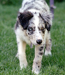
The Merle factor is a piebald gene that, if homozygous , can lead to malformations of the eyes such as the lack of a lens or reduced eyeballs in animals. It is encoded by the dog's silver locus. As with the horse's wind color gene, the merle factor only influences eumelanin.
Silver locus in horses: wind colors
In domestic horses , the silver gene corresponds to the wind color gene (silver dapple gene). The mutation is inherited as an autosomal dominant trait. She lightens the black mane from brown to white and the black fur from black horse to chocolate brown with light long hair. It has little or no effect on pheomelanin, so it does not lighten the fox-red color of completely brown horses.
Mutations of the gene in chickens: Dominant white, Dun and Smoky
Three mutations of the genetic locus discussed here are known in chickens: Dominant white, Dun and Smoky. The locus, which is called the silver locus in chickens, has nothing to do with the locus discussed here.
Dominant white and dun suppress the production of eumelanin, but allow the production of pheomelanin.
Dominant white: In this mutation, the transmembrane domain of the gene product is mutated. Chickens that are homozygous for the dominant white color, such as Leghorns, are white with brown eyes, as the mutation only affects the melanocytes from the neural crest. Despite the name, the gene is not completely dominant. Some of the heterozygous animals have black spots and most of the heterozygous animals that are not completely white had red color in their plumage (pheomelanin).
In addition to the dominant white mutation, Smoky has a second mutation that partially restores melanin production. This leads to a dark gray color. The mutation is partially dominant compared to wild type but recessive compared to dominant white
Dun is also caused by a mutation in the transmembrane domain that deleted six bases in the genetic code. Homozygous animals are almost white, while heterozygous animals are pale.
See also
- Coat colors of horses
- Genetics of horse colors
- Domestic rabbit genetics
- Coat colors of the cat
- Coat color
- Albinism
- Leucism
literature
- BS Kwon, R. Halaban, S. Ponnazhagan, K. Kim, C. Chintamaneni, D. Bennett, RT Pickard: Mouse silver mutation is caused by a single base insertion in the putative cytoplasmic domain of Pmel 17. In: Nucleic Acids Res . 23 (1), 1995 January 11, pp 154-158. PMID 7870580
- F. Solano, M. Martinez-Esparza, C. Jimenez-Cervantes, SP Hill, JA Lozano, JC Garcia-Borron: New insights on the structure of the mouse silver locus and on the function of the silver protein. In: Pigment Cell Res. 2000; 13 Suppl 8, pp. 118-124. PMID 11041368
- ZH Lee, L. Hou, G. Moellmann, E. Kuklinska, K. Antol, M. Fraser, R. Halaban, BS Kwon: Characterization and subcellular localization of human Pmel 17 / silver, a 110-kDa (pre) melanosomal membrane protein associated with 5,6, -dihydroxyindole-2-carboxylic acid (DHICA) converting activity. In: J Invest Dermatol. 106 (4), 1996 Apr, pp. 605-610. PMID 8617992
- T. Kobayashi, K. Urabe, SJ Orlow, K. Higashi, G. Imokawa, BS Kwon, B. Potterf, VJ Hearing: The Pmel 17 / silver locus protein. Characterization and investigation of its melanogenic function. In: J Biol Chem . 269 (46), 1994 Nov 18, pp. 29198-29205. PMID 7961886
- Emma Brunberg, Leif Andersson, Gus Cothran, Kaj Sandberg, Sofia Mikko, Gabriella Lindgren: A missense mutation in PMEL17 is associated with the Silver coat color in the horse. In: BMC Genet. 7, 2006, p. 46. doi: 10.1186 / 1471-2156-7-46 , PMID 1617113
- Emma Brunberg: Mapping of the silver coat color locus in the horse. Institutions for husdjursgenetik Sveriges Landbruksuniversitet, Uppsala 2006.
- LA Clark, JM Wahl, CA Rees, KE Murphy: Retrotransposon insertion in SILV is responsible for merle patterning of the domestic dog . In: PNAS . January 31, 103 (5), 2006, pp. 1376-1381.
- LL Baxter, WJ Pavan: Pmel17 expression is Mitf-dependent and reveals cranial melanoblast migration during murine development. In: Gene Expr Patterns. 3 (6), 2003 Dec, pp. 703-707. PMID 14643677 .
- M. Reissmann, J. Bierwolf, GA Brockmann: Two SNPs in the SILV gene are associated with silver coat color in ponies. In: Anim Genet. 38 (1), 2007 Feb, pp. 1-6. PMID 17257181
- Alexander C. Theos, Joanne F. Berson, Sarah C. Theos, Kathryn E. Herman, Dawn C. Harper, Danièle Tenza, Elena V. Sviderskaya, M. Lynn Lamoreux, Dorothy C. Bennett, Graça Raposo, Michael S. Marks : Dual Loss of ER Export and Endocytic Signals with Altered Melanosome Morphology in the silver Mutation of Pmel17. In: Mol Biol Cell. 17 (8), 2006 August, pp. 3598-3612. doi: 10.1091 / mbc.E06-01-0081 .
- Susanne Kerje, Preety Sharma, Ulrika Gunnarsson, Hyun Kim, Sonchita Bagchi, Robert Fredriksson, Karin Schutz, Per Jensen, Gunnar von Heijne, Ron Okimoto, Leif Andersson: The Dominant white, Dun and Smoky Color Variants in Chicken Are Associated With Insertion / Deletion Polymorphisms in the PMEL17 Genes. In: Genetics. 168, November 2004, pp. 1507-1518. doi: 10.1534 / genetics.104.027995

