Vertebrate eye

The vertebrate eyes are light-sensitive, often highly developed sensory organs , which sometimes serve to receive and transmit light stimuli in different ways ( eye ). Depending on the particular way of life and its requirements for visual orientation, this results in a more or less pronounced visual acuity , a differently sized peripheral field of vision and the varying perception of colors , shapes and movement .
In terms of evolutionary history , the eyes of vertebrates belong to the group of lenticular eyes and are very similar to one another, whereby their structure, with a few exceptions, corresponds in principle to that of the human eye. They are protected and embedded in a cushion of muscle, fat and connective tissue in the bony eye sockets ( orbita ) of the skull. Except for most fish, protective mechanisms such as the blink reflex prevent the eye from being damaged by external influences. The quick closing of the eyelid protects against foreign bodies on the one hand, and on the other hand prevents the sensitive cornea from drying out by constantly wetting it with tear fluid.
The movements of the eyes are controlled by the external muscles of the eye . The size of the resulting fields of vision is also very different here among the various genres and depending on the living conditions.
construction
The organ of vision (organon visus) of vertebrates is divided into three sub-units:
- the eyeball - Bulbus oculi (lat.) or ophthalmos (Greek),
- the appendix organs of the eye ( lacrimal system , eye muscles , conjunctiva and eyelids ) and
- the visual pathway .
eyeball

1 dermis (sclera)
2 choroid ( chorioidea )
3 Schlemm's canal ( sinus / plexus venosus sclerae )
4 oris root ( margo ciliaris )
5 cornea ( cornea )
6 iris ( iris )
7 pupil ( pupilla )
8 anterior eye chamber ( camera anterior bulbi )
9 posterior eye chamber ( camera posterior bulbi )
10 ciliary body ( corpus ciliare )
11 lens ( lens )
12 vitreous body ( corpus vitreum )
13 retina ( retina )
14 optic nerve ( nervus opticus )
15 zonular fibers ( fibrae zonulares )
Outer skin of the eye ( tunica externa bulbi ): 1 + 5
Middle eye skin ( tunica media bulbi / uvea ): 2 + 6 + 10
Inner eye skin ( tunica interna bulbi ): 13
The eyeball (synonym: Bulbus oculi ) is an almost spherical body that can rotate around any number of axes within certain limits, similar to the principle of a cardanic suspension , but does not change its position within the eye socket or only changes it insignificantly. Its shell consists of three concentric layers with different functions.
The interior of the eyeball, including the vitreous (vitreous body) , and the lens (lens) and is divided into front and rear chamber of the eye ( camera anterior and posterior bulbi ).
Outer eye skin
The outer skin of the eye ( tunica externa bulbi , also tunica fibrosa bulbi ) is divided into two sections. The transparent cornea is located where the light enters the eye . It is constantly moistened with tear fluid . It goes directly into the white dermis (sclera) , which forms the larger remaining part of the outer eyeball envelope. The outer eye muscles that move the eye in the eye socket are attached to it. In the anterior segment it is of the conjunctiva (conjunctiva) covered, so that only the cornea of tear fluid is wetted directly.
Middle eye skin
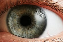
The middle skin of the eye ( tunica media bulbi or uvea ) consists of three sections. The choroid (choroidal) is rich in blood vessels , the adjacent layers supplied with nutrients and oxygen and is often pigmented. To the front, the choroid merges into the ciliary body (also known as the radiating body , corpus ciliare ), which is used to suspend and accommodate the lens of the eye . The foremost portion of the central eye skin is the iris (Iris). It forms the pupil and regulates the incidence of light ( adaptation ). Their pigmentation is what causes eye color .
Inner eye skin
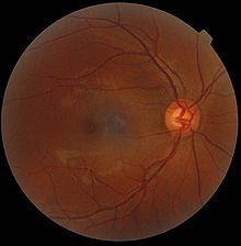
The inner eyes skin (Tunica interna bulbi) is of the retina (retinal) formed. This contains the light sensory cells ( photoreceptors ). There are no light-sensing cells where the optic nerve leaves the eye (optic nerve papilla). The area of the field of vision that corresponds to this point is called the blind spot . The point of sharpest vision is the fovea centralis , which is located within the yellow spot (macula lutea) . A pigment layer, the pigment epithelium, also belongs to the inner skin of the eye.
Vitreous
The vitreous body (corpus vitreum) is a gel-like, transparent substance that maintains the spherical shape of the eye. It is located inside the eye between the lens and retina and is part of the refractive media with a low optical effect . The vitreous body consists of approx. 98% water and approx. 2% hyaluronic acid and a network of collagen fibers (<< 1%).
Breaking media
In order to bundle the light that enters the eye from outside and focus it on the retina, optically effective components are required that ensure appropriate light refraction . These are summarized under the term “refractive media” and consist of the cornea , the lens , the aqueous humor and the vitreous humor . Their share in the total refractive power differs - also from living being to living being. In principle, however, the cornea has by far the greatest refractive power , followed by the lens. The totality of the refractive media is also known as the dioptric apparatus , and their refractive power is given in the unit of diopters (dpt).
Annex organs
The appendix organs of the eye include the lacrimal system , the eye muscles , the conjunctiva and the eyelids .
Lacrimal system
The tear system of land-living vertebrates consists on the one hand of the structures that are responsible for the production of tear fluid ( lacrimal gland ), on the other hand of the supply and drainage vessels and ducts ( tear ducts ) that transport the tear fluid. The entire organ is used to supply the anterior sections of the eye, to clean them and to protect them. The tear fluid is drained through a drain in the corner of the eye and finally flows into the nasal cavity via the tear and nasal duct .
External eye muscles
The vertebrate eye has seven external eye muscles (six in humans). They are divided into four straight and two oblique eye muscles, each of which can turn each eye in different directions and, in pairs, exert antagonistic forces on the eyeball . Depending on the position of the eyes, the muscles have more or less pronounced main and partial functions, which are expressed in the raising, lowering, sideways turning or rolling of the eyeball. Many mammals also have one other muscle that has a similar range of functions to the four straight muscles.
The eye movements triggered in this way occur on the one hand with the aim of aligning the lines of the face with an object to be fixed in the outside space, and this is coordinated as precisely as possible and in the shortest possible time. In living beings with central fixation , the fovea centralis not only represents the main direction of vision but also the motor zero point of the eye with regard to its movement physiology and thus enables not only a subjective localization in space but also spatial vision . On the other hand, the eye movements enlarge the field of vision . The monocular excursion route is important, i.e. the maximum mobility of the right and left eye, which can be very different from living being to living being. The performance of the eye muscles is usually many times higher than is actually required on a daily basis.
Conjunctiva
The conjunctiva, also known as the conjunctiva , is a mucous membrane in the orbit (eye socket) in the anterior segment of the eye. It begins at the edge of the eyelid and, as a tunica conjunctiva palpebrarum, covers the rear surface of the eyelids facing the eyeball . This mucous membrane coating acts like a soft wipe and, when you blink, distributes the tear fluid over the cornea without damaging it. In the depth of the eye socket, the conjunctiva folds forward again and connects with the sclera . The conjunctiva covers the front part of the sclera up to the beginning of the cornea; this section is called the tunica conjunctiva bulbi .
The cavity enclosed by the conjunctiva is the conjunctival sac ( Saccus conjunctivae ). Its rear niche in the depths of the eye socket is called the conjunctival vault ( Fornix conjunctivae ).
The conjunctiva forms an additional fold on the nasal corner of the eye, which is called the nictitating membrane ( Plica semilunaris conjunctivae , Membrana nicitans ) or third eyelid ( Palpebra tertia ). It is only very small in humans. In the other mammals , it is so large that it can lie in front of the entire eye. In many other vertebrates , e.g. B. sharks , reptiles and birds , it is transparent and can be folded over the eye like protective goggles.
Eyelids
The eyelid is a thin fold consisting of muscles , connective tissue and skin , which an eye can completely cover in order to protect it from external influences and foreign bodies, among other things by means of a reflex ( eyelid-closing reflex ). With every blink of the eye , it distributes tear fluid , which is deposited in the form of a tear film over the front surface of the eyeball, thus keeping the sensitive cornea clean and moist.
There is an upper ( superior palpebra ) and a lower eyelid ( inferior palpebra ). The eyelid gap ( rima palpebrarum ) is located between the two . Both eyelids meet on the sides in the corner of the eyelid ( angulus oculi or canthus ). Many vertebrates also have eyelashes on the upper and lower eyelids, which also protect the eyes.
Visual pathway
All transmission lines and neural connections of the optical system from the eye to the brain are called the visual pathway. These include the retina in the eye, the optic nerve up to its course at the optic nerve junction and the adjoining optic tract . In the lateral knee hump of the diencephalon ( corpus geniculatum laterale ) , the first interconnections of the visual pathway outside the retina take place. It continues as so-called Gratioletian visual radiation to the primary visual cortex .
Retina
The retina ( retinal ) is a light-sensitive structure of nerve tissue at the rear and laterally inward side of the eye. In it the incident light is converted into nerve impulses. In addition to the light-sensitive tissue, the retina consists of nerve cells for processing and forwarding the generated impulses, as well as various support structures for maintaining the function of stimulus-generating and stimulus-processing cells.
Optic nerve
The optic nerve ( Nervus opticus ) is the second cranial nerve and after the retina the first section of the visual pathway . It enters the eyeball on the optic nerve head, the papilla, and is on average 4.5 cm long. The optic nerve is a route of bundled nerve fibers from the sieve plate (lamina cribrosa) of the dermis of the eye to the junction of the optic nerve (chiasma opticum) and can be divided into three parts:
- an intrabulbar part located in the eyeball
- an intraorbital part located within the eye socket ( orbit )
- an intracranial part located in the skull
The optic nerve contains around a million nerve fibers, the projections ( axons ) of the ganglion cells of the retina ( retina ). The nasal half of the fibers, which transport the signals from the nasal half of the retina, crosses in the optic nerve junction ( optic chiasm ) to the optic tract on the other side, so that the signals from the left visual field reach the right half of the brain and vice versa. From the point of entry into the optic nerve, the nerve fibers are individually surrounded by the myelin sheaths of the oligodendrocytes , which lead to an increased conduction velocity, but prevent regeneration if damaged. Numerous astrocytes are also found in the vicinity of the axons.
Optic tract
The optic tract continues from the optic chiasm as the optic tract . Most of the information reaches the corpus geniculatum laterale (CGL, lateral knee hump) of the diencephalon .
Mechanism of image generation
The light reaches the interior through the cornea and the pupil . It is the circular opening of the colored iris, the iris . The muscle fibers in the iris can enlarge and reduce the size of the pupil. This process, which adapts the eye to the brightness of the surroundings, is called adaptation . The elastic lens of the eye is suspended from ribbons behind the iris . The lens bands run to the ring-shaped ciliary muscle . The inside of the eye is filled with the gelatinous glass body . It gives the eye the firm and round shape, which is also called the eyeball.
The area of the electromagnetic spectrum that can be perceived by the eye ( light spectrum ) differs from living being to living being. The visual space in the vertebrate eye is much smaller for colored vision (colored light, through the cones ) than that for light and dark (white light, through the rods ). Thus, the color space is also within the white light space.
Although most of the retina (pars optica retinae) is covered with sensory cells , sharp vision only occurs within certain limits, the so-called yellow spot ( macula lutea ). In principle, only the area that the eyes fix with their facial lines is seen in focus .
When looking at an object, a constant and sharp image only comes about when the eye muscles , mostly unconsciously, move various sections of the object one after the other over an area of about 1.0 ° in front of the fovea centralis , the center of the yellow spot. The eye never rests when looking at it and is always involved in the smallest of movements by fixing a point for fractions of a second and then following a smallest, jerky movement ( microsaccade ) to the next point. The overall image is finally generated from this “scanning”. When viewed calmly, the individual fixations last 0.2 to 0.6 seconds, so that 2 to 5 saccades take place in one second. If you look more quickly, the saccades become more frequent and the fixation times shorter.
The choice of the fixation points and the pattern of the saccades is highly individual and is related to the habits and interest of the observer or the task at hand. Today we speak of “intentional seeing”, an active process towards the world. By means of appropriate observation methods, the advertising industry in particular , but also behavioral research, make use of this phenomenon of involuntary activity in order to improve and optimize their advertising methods and their theses on human behavior. In connection with the development of lie detectors , appropriate instruments are often used, mostly to assess the state of arousal.
Accommodation
Most vertebrates have the ability to use a variety of mechanisms to view objects that are at different distances. This process is called accommodation, which means something like "adaptation". A distinction is made between near accommodation , in which the eyes adjust to objects close by, and distant accommodation , the adjustment to things that are at a greater distance. While the eyes of mammals, amphibians and also sharks are set to the distance in a relaxed state and accommodation is usually close to, the eyes of other aquatic vertebrates are set to close sight, and an adjustment only takes place for the distance. One differentiates between the mechanisms underlying the adjustment processes
- static systems in which the optical variability is achieved through structural features, as well as
- dynamic systems which are based on an active change in the dioptric apparatus through muscle strength.
In humans, the accommodation decreases with increasing age, which ultimately leads to presbyopia , which can be corrected with appropriate aids, such as glasses .
The human eye
properties
The sense of sight is very important to humans. He is the guiding sense that enables him and other visually oriented living beings a safe orientation. In practical terms, this is also expressed in the amounts of compensation that insurance companies pay for the loss of one or both eyes . In Germany, the degree of disability in the event of loss of an eye is given as 50 percent.
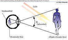
The appropriate stimulus for the eye , as a sensory organ, is created in humans by electromagnetic radiation with a wavelength between about 400 and 760 nanometers and is somewhat different for day and night vision (see sensitivity curve ). The anatomical and functional structure of the eyeball ensures that the central property of the human sense of sight, visual acuity , is of a correspondingly high quality. It arises in an approximately 5 ° area of our binocular field of view, which encompasses around 170 ° horizontally and around 110 ° vertically .
At birth, the eye does not have full vision. Only in the course of the first few months of life do newborns learn to fixate on things in their environment and thus to provide the necessary stimulus that the visual system needs for adequate development of visual acuity. In early childhood, the eyes usually have a physiological farsightedness of +2.0 to +3.0 diopters. Due to the anatomical growth, the optical conditions also change. The farsightedness is therefore ideally reduced to around +0.5 diopters up to adulthood.
The human eye belongs to the group of lens eyes. The optical system required to refract light, the dioptric apparatus , has a total refractive power of around 60 diopters ( Emmetropauge according to Gullstrand 58.64 dpt). The respective optically active components cornea , lens , aqueous humor and vitreous , the so-called refractive media , have different proportions. The entire system ensures that the light rays entering the eye are focused at the point of sharpest vision, the fovea centralis . Through the process of accommodation , this is possible in the most varied of distances between the optical far and near point.
Even if it appears as if the human eye fixes things in the outside space calmly and motionlessly, it nonetheless performs one to three very small gaze leaps per second, so-called microsaccades . This prevents over-stimulation of the sensory cells on the retina, which is called local adaptation .
The color of the eyes is caused by different pigmentation of the iris . Due to the storage of the brown-coloring melanin in the iris own layer, a characteristic eye color is formed which, depending on the amount of pigment, ranges from gray, yellow, green to brown, and with a correspondingly high amount of melanin even to black. In humans, this mostly correlates with skin and hair color. Light-skinned and blonde people tend to have blue eyes, while dark-skinned people with dark hair usually have a brown iris color. Around 90 percent of all people worldwide have brown eyes, including the vast majority of people of non-European descent. The rest is divided into blue, green and gray. According to the theory of genetic researcher Hans Eiberg of the University of Copenhagen, all blue-eyed people should be descended from one and the same human.
While in many other living beings the two or more eyes are used exclusively to enlarge the field of vision and field of vision, the human sense of sight is also clearly designed for binocularity , i.e. a fusion of the visual impressions of the right and left eye. Only this ability, as a result of precise coordination and cooperation, enables high-quality spatial vision . In contrast, the quality of the visual acuity is only mediocre in comparison, for example, with that of birds of prey .
The human eye in numbers
All values are average values for emmetropia and can vary according to gender and age.
| eyeball | tears | Dermis | Choroid | ||||
| ø in the newborn | 17 mm | Start of tear production | approx. 3rd week of life | Thickness of the dermis
(behind the rectus muscle) |
0.3 mm | Thickness in the macular area | 0.22-0.30 mm |
| ø in adults | 24 mm | Production amount children | 84 mg / h | Thickness of the dermis
(Near the optic nerve) |
1.35 mm | Thickness in the equatorial area | 0.10-0.15 mm |
| Weight | 7.5 g | Production volume adults | 38 mg / h | Distance of the capillary mesh in the macular area | 3-18 µm | ||
| volume | 6.5 cm³ | Total protein | 6.69 g / l | Distance of the capillary mesh in the equatorial area | 6-36 µm | ||
| density | 1.002-1.090 g / ml | Total albumin | 3.94 g / l | ||||
| scope | 74.9 mm | Total globulin | 2.75 g / l | ||||
| Intraocular pressure | 10-21 mmHg | Daily production quantity | 1–500 ml (1 ml = approx. 1 g) | ||||
| Cornea | iris | Aqueous humor | lens | ||||
| thickness | 0.52-0.67 mm | diameter | 12 mm | Production quantity | 2 mm³ / min | Thickness at
Newborn |
3.5 mm |
| surface | 1.3 cm² | Thickness in the area
the orris root |
0.5 mm | Daily exchange rate | 2-3 ml | Thickness in child
at the age of 10 |
3.9 mm |
| ø vertical | 10.6 mm | Thickness in the area
the iris frill |
3.0 mm | protein | 6.69 g / l | Thickness for adults
20–50 years |
4.0-4.14 mm |
| ø horizontal | 11.7 mm | Pupil diameter (adaptation) | 1.2-9.0 mm | Table salt | 6.58 g / l | Thickness for adults
60–70 years |
4.77 mm |
| Refractive power | 43 dpt | sodium | 4.45 g / l | Thickness for adults
80–90 years |
5.0 mm | ||
| Refractive index | 1.34 | potassium | 1.16 g / l | Lens diameter | 6.5-9 mm | ||
| Glucose | 0.65 g / l | Thickness of the lens capsule at the posterior pole | 2-4 µm | ||||
| Refractive power | 19–33 dpt | ||||||
| Weight | 1.74 g | ||||||
| Vitreous | Retina | Optic nerve | |||
| volume | 4 cm³ | Thickness at the equator | 0.18 mm | ø intraorbital section | 3-4 mm |
| Weight | 4 g | Thickness in the fovea centralis | 0.10 mm | ø intracranial section | 4-7 mm |
| Refractive index | 1.334 | Thickness at the optic nerve | 0.56 mm | Number of nerve fibers in the optic nerve | 1,000,000 |
| ø macula, vertical | 0.88 mm | Intraocular length | 1 mm | ||
| ø macula, horizontal | 2.0 mm | Length intraorbital | 25 mm | ||
| Number of chopsticks | 125,000,000 | Intra-canalicular length | 4-20 mm | ||
| Number of cones | 7,000,000 | Intracranial length | 10 mm | ||
| Number of retinal switching cells | 2,000,000 | ||||
| Visual acuity , sensitivity | binocular, horizontal field of view |
Accommodation near point / accommodation width |
Oculomotor function | ||||
| Resolving power | 1/120 arc degrees | 16-19 years | 174 ° | 10–19 years | 7 cm / 14 dpt | Excursion routes | Abduction / Adduction: 50 ° Elevation: 60 ° Lowering: 45 ° |
| Smallest viewing angle | 20 ″ | 20-29 years | 175 ° | 20-29 years | 9 cm / 11 dpt | Saccades | Speed: 600 ° / sec Number: 1–3 / sec |
| Angle of view that corresponds to the size of a cone | 0.4 ″ | 30–39 years | 174 ° | 30–39 years | 12 cm / 8 dpt | Follow-up movements | Speed: 100 ° / sec |
| Distance on the retina corresponding to 1 ° | 0.29 mm | 40-49 years | 172 ° | 40-49 years | 22 cm / 4.5 dpt | ø muscle pull | 0.1-0.5 N |
| Lower awareness threshold | 1–2 arc minutes / sec | 50–59 years | 167 ° | 50–59 years | 40 cm / 2.5 dpt | Basic muscle tone | 0.05-0.1 N |
| Perception of movement and direction | 300-400 ° / sec | 60–69 years | 160 ° | 60–69 years | 100 cm / 1 dpt | Maximum muscle pull | 1 N |
| Perception of movement alone | from 600 ° / sec | 70–79 years | 151 ° | > 70 years | up to 400 cm / 0.25 dpt |
pivot point | 13.5 mm behind the vertex of the cornea |
| Absorption area of the photoreceptors (violet - red) |
400-760 nm | > 80 years | 140 ° | ||||
| Sensitivity of maximum light adaptation and full dark adaptation | 1:10 6 | ||||||
| Minimum number of photons that excite a rod | 5 | ||||||
| Absolute stimulus threshold for twilight vision | 2–6 × 10 −17 Ws | ||||||
| Temporal resolution | 60-65 Hz | ||||||
| Duration of adaptation to darkness | 30 minutes | ||||||
Tear fluid has about as much salt as blood plasma, i.e. approx. 0.9%, and has a slightly basic reaction: pH = 7.35. The tears come (per eye) from the bean-sized lacrimal gland, which lies below the outside of the eyebrow, flows through 6 to 12 tear ducts on the upper eyeball and is distributed over the cornea with every blink of the eye. The liquid forms a layer of mucus on the eye, it is watery but the surface is fatty and hydrophobic in order to reduce the evaporation of water. Excess tear fluid flows through 2 tear ducts, small holes visible in the inner corner of the eyelid, into the tear sac on the side of the nose and further into the nose. Tear production is increased in the case of strong emotions (crying, laughing, joy, sadness) or chemical or physical stimuli such as smoke or cold, but it is reduced during sleep.
Diseases and dysfunction
→ See also: Diseases in ophthalmology
The Ophthalmology deals with the prevention , diagnosis and treatment of eye diseases. These lead primarily to a more or less pronounced loss of functional performance, such as a reduction in visual acuity , restrictions in the field of vision, color vision disorders , reduction in twilight vision or disorders of binocular vision. In addition, viral and bacterial infections as well as local inflammatory processes, injuries, pain , swelling, tumor formation , increased tearing, increased sensitivity to glare and movement disorders are other possible symptom complexes and organic impairments.
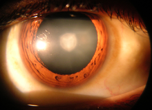
The most common diseases with a visual acuity reduction next to include cataracts ( cataract ) and glaucoma ( glaucoma ) age-related macular degeneration and diabetic retinopathy . The most common forms of cataract, glaucoma and macular degeneration are thought to be caused by age changes based on genetic predisposition. For macular degeneration in particular, smoking is the essential exogenous risk factor. It is also suspected that ultraviolet light has a harmful effect on cataracts and macular degeneration . Diabetic retinopathy is the result of changes in the blood vessels caused by the increased blood sugar level. It also occurs earlier and more frequently in smokers than in non-smokers.
The healing prospects with the conservative and surgical treatment options available are very different. Diseases with involvement of the retina often have a less favorable prognosis because it is neither regenerative nor permanently replaceable. This can lead to detachments ( amotio ), holes (foramen), tears ( rupture ) or splitting of retinal layers ( retinoschisis ). In the case of some retinal diseases (e.g. retinopathia pigmentosa ), it is hoped that in the future the visual function will be restored with a retinal implant .

Furthermore, inflammatory processes are often found, especially on the conjunctiva ( conjunctivitis ), the cornea ( keratitis ), the iris ( iritis ) and the choroid ( uveitis ), but also on the lacrimal system and the lids ( blepharitis ). Pathological cloudiness can also occur within the vitreous humor .
A functional disease of the eye without a recognizable organic cause in the form of a sometimes massive reduction in visual acuity is called amblyopia . It is caused, among other things, by certain strabismus diseases or very different refraction ratios ( anisometropia ).
Reductions in the image quality on the retina and thus in visual acuity can be caused by refractive defects ( ametropia ) such as myopia , farsightedness or astigmatism . With age, the lens also loses its elasticity, which reduces the ability to focus closely and leads to presbyopia .
There is a whole range of diseases that have their causes in a completely different place, but nevertheless manifest themselves with corresponding symptoms on or in the eye. These include, in particular, certain neurological diseases, endocrine orbitopathy as an expression of a hormone- related autoimmune process , diabetes mellitus , circulatory and metabolic disorders , toxoplasmosis or multiple sclerosis . For this reason, ophthalmological diagnostics play an important role in the identification of interdisciplinary clinical pictures.
The eye is often exposed to external influences that can lead to injuries, for example from foreign bodies, blunt contusions (punch, tennis ball, etc.) or flashes .
Nonspecific visual impairments are also summarized under the term visual impairment .
Investigation procedure
→ See also: Diagnostic procedures in ophthalmology and diagnostic tools in ophthalmology
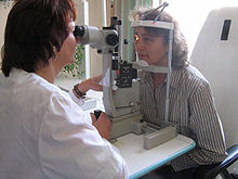
Ophthalmological diagnostics is characterized by a large number of apparatus-based examination procedures and, in the case of organic assessment, primarily extends to the inspection of the visible components of the anterior, middle and posterior segments of the eye. Special devices are used with which almost all organ areas can be viewed and assessed. One of the most important here is the slit lamp , which, partly in combination with other aids such as the contact glass , enables all essential sections to be assessed. In conjunction with an applanation tonometer , intraocular pressure can also be measured. More compact variants, especially for assessing the fundus, are found in what are known as ophthalmoscopes . Investigations of the fundus are often prepared with medication to enlarge the pupil ( Mydriatica ) in order to provide a better insight.
The measurements and investigations of the functionality of the sense of sight are also carried out with a range of aids and apparatus. Phoropters and refractometers , for example, are used to test visual acuity ( eye test ) and the optical conditions of the eyes . The field of vision is measured with a perimeter , twilight vision and glare sensitivity are determined by a nyctometer , and the color sense is checked with the so-called Ishihara color tables , among other things . The investigation of binocular vision and the mobility of both eyes has even spawned an independent specialty . There are inspection and test procedures for all functions of the human sense of sight that provide reproducible results and thus enable detailed progress documentation. Various methods of ophthalmic electrodiagnostics (ENG, ERG, VEP etc.) enable the assessment of movement sequences, the sensitivity of the retina and the visual pathway.
Also include imaging techniques such as optical coherence tomography (OCT), or the Heidelberg retina tomograph (HRT) is now the diagnostic standard in ophthalmology.
Eyes in different groups of vertebrates
Mammals
The functional performance of the eyes and thus the quality of visual perception is sometimes very different in mammals and depends on the respective living conditions. In living beings that live underground (e.g. moles ), the eyes are often regressed, while cats and other predators have a powerful fovea centralis and have correspondingly good eyesight. As with humans, the position of their eyes is also geared towards differentiated spatial vision. Escape animals such as horses or typical prey such as rabbits , on the other hand, have a lateral arrangement of the eyes that does not allow spatial vision, but enables a large field of vision and has high density “visual stripes” of retinal sensory cells that are parallel to the horizon.
Birds
The eyes of birds are larger in relation to their body size than those of mammals. Some of them have special functional adaptations to their environment.
The eyes of nocturnal birds ( e.g. owls ) with less visual acuity allow a higher light output than those of day birds (e.g. peregrine falcons ). Some small birds are also able to see UV light . In contrast to humans, many species have four instead of three color receptors . Most bird species can also perceive more images per second than humans and thus achieve a higher temporal resolution . The retinal area of sharpest vision is around 20 ° in birds, around eight times as large as in humans. Diurnal birds of prey also have two foveae, a medial one for binocular fixation and a lateral one for lateral perception.
Reptiles
Reptile eyes can vary widely in size and in some cases be significantly reduced. They are usually located in large eye sockets and are usually moved by the six outer eye muscles and the retractor bulbi muscle.
The appendage organs partly show lacrimal glands, partly they are missing. However, all species have so-called Harder's glands , which produce a secretion that enables the nictitating membrane to glide over the eyeball. While the secretion of snakes and some types of lizards , in which the lower eyelid has grown into a transparent, rigid membrane ("glasses"), is drained into the oral cavity via lacrimal ducts, in crocodiles it can , for example, with strong swallowing movements, between the nictitating membrane and Eject the eyeball and lead to the well-known crocodile tears.
The type and number of stimulus-processing retinal cells (cones and rods) and the shape of the pupils (round or slit pupils) depend on the day or night activities of the respective species. Lizards have two to three different types of cones, and more highly developed snakes have additional rods.
Amphibians

The eyes of amphibians , with the exception of species that live underground or cave-dwelling, are usually well developed. Except for the tail and frog grooves, they have movable eyelids. The retractor bulbi muscle still has the distinctive function of pulling the eyeball back into the orbit, especially when catching prey and when swallowing.
The retina has two types of rods and cones. The field of vision can be very large with a dimension of up to 360 ° ( frogs and some salamander species ) and allow an almost complete all-round view. The possibility of spatial vision is provided by appropriate overlaps .
Aquatic vertebrates
The eyes of aquatic vertebrates show some anatomical and functional features that distinguish them from those of other vertebrates.
While the eyes of the majority of ray fins , the hagfish and some electric rays are covered by a transparent layer of skin and the cornea of the sharks is protected by a translucent nictitating membrane , other fish usually lack eyelids or similar protective mechanisms. The eyes of the hagfish also have neither a lens nor an iris. Cartilage and bony fish, on the other hand, have iris and a lens, which, however, are inelastic and focused on near vision. However, it can be changed in position by muscle contraction and thus adapted to a distance view.
literature
- Simon Ings : The eye. Masterpiece of evolution. Hoffmann and Campe, Hamburg 2008, ISBN 978-3-455-50072-1 .
- Theodor Axenfeld (founder), Hans Pau (ed.): Textbook and atlas of ophthalmology. With the collaboration of Rudolf Sachsenweger a . a. 12th, completely revised edition. Gustav Fischer, Stuttgart a. a. 1980, ISBN 3-437-00255-4 .
- Winfried Ahne, Hans-Georg Liebich, Manfred Stohrer, Horst Erich König, Eckhard Wolf: Zoology. Textbook for students of veterinary medicine and agricultural sciences. Schattauer, Stuttgart 2000, ISBN 3-7945-1764-4 .
Web links
- LP - The human eye as an optical system (Georg-August-Universität Göttingen)
- Light sensory cells - construction and photoreception teaching material (PDF; 513 kB)
- Katja Seefeldt: Where Darwin Still Shuddered - Origins of the Human Eye (Telepolis, October 31, 2004)
- Trevor D. Lamb: Evolution of phototransduction, vertebrate photoreceptors and retina - from sciencedirect.com, June 18, 2013
Individual evidence
- ↑ a b c d e f Theodor Axenfeld (founder), Hans Pau (ed.): Textbook and atlas of ophthalmology. With the collaboration of Rudolf Sachsenweger a . a. 12th, completely revised edition. Gustav Fischer, Stuttgart a. a. 1980, ISBN 3-437-00255-4 .
- ↑ a b c d e f g h Herbert Kaufmann (Ed.): Strabismus. 3rd, fundamentally revised and expanded edition. Georg Thieme, Stuttgart a. a. 2003, ISBN 3-13-129723-9 .
- ^ Erwin Deutsch: Insurance contract law. A floor plan. 5th, revised edition. Verlag Versicherungswirtschaft, Karlsruhe 2005, ISBN 3-89952-177-3 .
- ↑ Peter Frost: Why Do Europeans Have So Many Hair and Eye Colors? ( Memento from January 2, 2008 in the Internet Archive ) Yet skin color is weakly influenced by the different alleles for hair color or eye color, apart from the ones for red hair or blue eyes. Some have no effect at all on skin pigmentation.
- ↑ David L. Duffy, Neil F. Box, Wei Chen, James S. Palmer, Grant W. Montgomery, Michael R. James, Nicholas K. Hayward, Nicholas G. Martin, Richard A. Sturm: Interactive effects of MC1R and OCA2 on melanoma risk phenotypes. In: Human Molecular Genetics. Vol. 13, No. 4, January 2004, ISSN 0964-6906 , pp. 447-461, doi: 10.1093 / hmg / ddh043 . All blue-eyed R / R individuals (Note: R stands for a strong red-haired allele, in contrast to r. Both are recessive.) Were in the fair / pale skin category but this decreased to 85.4% with fair / pale skin for brown / green-eyed R / R individuals, the remainder having medium skin color. This proportionate lightening in all genotypic groups when carrying both recessive blue-eyed b and red-hair R alleles indicates additive action of MC1R and BEY2 / OCA2 loci on constitutive skin color.
- ^ Hans Eiberg, Jesper Troelsen, Mette Nielsen, Annemette Mikkelsen, Jonas Mengel-From, Klaus W. Kjaer, Lars Hansen: Blue eye color in humans may be caused by a perfectly associated founder mutation in a regulatory element located within the HERC2 gene inhibiting OCA2 expression. In: . Vol. 123, No. 2, ISSN 0340-6717 , pp. 177-187, doi: 10.1007 / s00439-007-0460-x .
- ↑ The eye in numbers.
- ↑ Robert F. Schmidt , Florian Lang (ed.): Physiology of humans. With pathophysiology. 30th, revised and updated edition. Springer Medicine, Heidelberg 2007, ISBN 978-3-540-32908-4 .
- ^ Anatomy of the eye> tear fluid dr-gumpert.de, accessed on April 2, 2018.
- ↑ Dr. Constanze Fünfstück, Jena Eye Clinic: Expert from Jena explains how the salt gets into tears jena.otz.de, June 30, 2013, accessed on April 2, 2018.
- ↑ Leslie Hyman et al. a .: Prevalence and causes of visual impairment in the Barbados eye study. In: Ophthalmology. Vol. 108, No. 10, 2001, ISSN 0161-6420 , pp. 1751-1756.
- ↑ Hope for the blind: Electronic vision aid in prospect. ( Memento from July 20, 2006 in the Internet Archive )
- ↑ a b c Wilfried Westheide, Reinhard Rieger (Ed.): Special Zoology. Volume 2: Vertebrates or Skull Animals. Spectrum, Academic Publishing House, Heidelberg a. a. 2004, ISBN 3-8274-0900-4 , p. 84.
- ↑ Wilfried Westheide, Reinhard Rieger (Ed.): Special Zoology. Volume 2: Vertebrates or Skull Animals. Spectrum, Academic Publishing House, Heidelberg a. a. 2004, ISBN 3-8274-0900-4 , p. 387.
- ↑ Wilfried Westheide, Reinhard Rieger (Ed.): Special Zoology. Volume 2: Vertebrates or Skull Animals. Spectrum, Academic Publishing House, Heidelberg a. a. 2004, ISBN 3-8274-0900-4 , pp. 363-364.
- ↑ Wilfried Westheide, Reinhard Rieger (Ed.): Special Zoology. Volume 2: Vertebrates or Skull Animals. Spectrum, Academic Publishing House, Heidelberg a. a. 2004, ISBN 3-8274-0900-4 , p. 314.


