Perimetry
As perimetry (from the Greek peri "around", metron "dimension") is known in the ophthalmology , of Neurology and the optometry the systematic measurement of the visual field .
The aim of the investigation is on the one hand to determine the outer and inner limits of the visual field and on the other hand to determine the sensitivity of the visual system in the perceived area. The devices used here are called perimeters .
During the examination, optical stimuli are presented one after the other at different locations in the room. The perception of these stimuli is recorded depending on their location and their strength. In order to maintain the spatial relationship between the test sites, the examined eye must continuously fix a central point. A schematic image of the visual field can then be constructed from the examination protocol. The partner's eye must be covered with an eye patch, for example, throughout the entire examination process. As a rule, the results can only be used with good cooperation from the person examined.
A basic distinction must be made between static and kinetic investigation methods. In the former, the stimuli are presented at fixed locations and their intensity increased or decreased until the person examined signals a perception or no longer signals it. With the latter, stimuli that are invariable in their intensity are moved from outside the visual field limits into the assumed visual field and the location of the perception is viewed as the limit of the visual field for the given stimulus intensity.
The brightness (more precisely: luminance ) of the visual stimuli and the examination background are in the sensitivity range of the cones (photopic vision). Central scotoma , the fixation complicate the patient. However, they can be measured in terms of size and depth with both contour perimetry and threshold perimetry.
history
A device for measuring the field of view was already manufactured by Claudius Ptolemy around 150 AD. The modern perimeter was invented by the physiologist Hermann Aubert and reported on it in 1857. The ophthalmologist Richard Förster was then able to improve the device. This Förster perimeter , a circular arc perimeter, was presented at the ophthamology congress in Paris in 1867.
Investigation methods
Finger perimetry
Finger perimetry ( synonym: confrontation perimetry or confrontation field of view) is a kinetic method that has the advantage of making qualitative statements about the external limits of the field of view without extensive technical equipment. The examiner compares the outer limits of his own field of vision with those of the test subject. The examiner and the test subject sit opposite each other, each covering an opposite eye and mutually fixing the other's nose, for example. The examiner's finger is guided into the field of vision from the outside, and the subject explains when he notices the finger. Finger perimetry is only suitable for the determination of gross visual field defects, is prone to errors and does not provide any information about the distribution of sensitivity within the visual field.
Contour perimetry
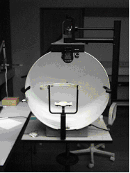
The contour perimetry ( synonym: Goldmann perimetry, isoptera perimetry) is the classic method of perimetry, also kinetic. The patient's head is in a projection perimeter (a hollow-sphere perimeter developed by Goldmann in the 1940s that replaced the arched perimeter). The test point is projected into this sphere and is mechanically coupled to a guide pin in such a way that the position of the test point is transferred to a flat sheet of paper. The size and brightness of the test point can be selected independently of one another. The result of the contour perimetry shows the field of view similar to a map with contour lines as curves of equal sensitivity (isopters).
Threshold perimetry (computer perimetry)
Threshold perimetry ( synonym: computer perimetry, static perimetry) is a procedure that determines the perception threshold at fixed points in the visual field . The test person looks into an optical system - usually also a hemisphere, over which points of light of different positions and brightness are projected computer-controlled. The test person confirms each recognized stimulus with the push of a button. With the help of so-called adaptive threshold determination methods, both the entire extent of the field of view and the condition of selected areas can be examined under various questions. The results can be processed directly for electronic documentation.
Before the introduction of computer-controlled threshold perimetry, static perimetry was performed manually as profile perimetry . As an examination device z. B. a perimeter of the Goldmann type, or a Tübingen perimeter according to Harms and Aulhorn, used (see contour perimetry). The light stimuli offered were not moved, but increased in intensity by means of a slider. Because of the effort involved, this examination was usually limited to only one or a few meridians of the visual field, so that the result was a profile of the visual field map.
See also
- glaucoma
- Retinopathia pigmentosa
- Hemianopia
- Quadrant anopia
- Light tail test
- Visual pathway
- Optic nerve
- Scotoma
- Peripheral vision
Web links
- DIN EN ISO 12866 - Ophthalmic instruments - Perimeter. DIN NA 027 Standards Committee Precision Mechanics and Optics (NAFuO)
literature
- Theodor Axenfeld (founder), Hans Pau (ed.): Textbook and atlas of ophthalmology. With the collaboration of Rudolf Sachsenweger a . a. 12th, completely revised edition. Gustav Fischer, Stuttgart a. a. 1980, ISBN 3-437-00255-4 , p. 45 ff.
Individual evidence
- ↑ Frank Krogmann: Perimeter (= device for measuring the field of view). In: Werner E. Gerabek , Bernhard D. Haage, Gundolf Keil , Wolfgang Wegner (eds.): Enzyklopädie Medizingeschichte. De Gruyter, Berlin / New York 2005, ISBN 3-11-015714-4 , p. 1121.
- ↑ Anselm Kampik, Franz Grehn (ed.): Ophthalmic therapy. Thieme, Stuttgart a. a. 2002, ISBN 3-13-128411-0 , p. 410.
- ↑ Wolfgang Leydhecker: Advances in modern ophthalmology. In: Würzburg medical history reports. Volume 3, 1985, pp. 185-210, here: pp. 198-200.
