Adaptation (eye)
In the case of the eye, adaptation ( lat. Adaptare "to adapt") is understood to mean its adaptation to the luminance prevailing in the visual field .
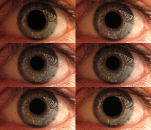
In the event of sudden differences in brightness, the amount of incident light can be quickly adjusted via the pupillary light reflex through narrowing or widening of the pupils with the iris musculature , but this only allows a change within a range of about 1:10.
The further adjustment to different ambient brightness is achieved by changing the light sensitivity of the retina and is only optimal after a certain delay. Only this retinal adaptation makes it possible to process light stimuli of very different strengths - for example, to see faintly shining stars in the moonless night sky or to see traces in the snow in sunlight (range about 1:10 12 ). In addition to biochemically adapted transduction processes in the sensory cells ( photoreceptors ), differently weighted interconnections within the receptive field of downstream retinal nerve cells also contribute to this.
Pupillary light reflex
The iris (iris) represents the delimitation of the eye hole ( pupil ). The result of the pupillary light reflex , or pupil reflex for short , is a change in the tone of the smooth iris muscles . This causes a change in the pupil size, whereby the relative amount of light entering the eye can be adjusted. The mechanism is comparable to regulating the aperture of a camera. The iris has two muscles for this:
- The dilator pupillae muscle ("pupil widener") is innervated by sympathetic nerve fibers from the centrum ciliospinale (spinal cord segments C8-Th3). The dilation of the pupil is known as mydriasis .
- The sphincter pupillae muscle ("pupil constriction") is made up of parasympathetic fibers of the oculomotor nerve (third cranial nerve ) from the Ncl. Edinger-Westphal is innervated via the ciliary ganglion . It is activated when there is high incidence of light. A narrowing of the pupil is known as miosis .
The reflective regulation of the incidence of light by the pupil causes a rapid adaptation to sudden changes in brightness. If the pupil diameter increases by three times, the enlargement of the opening area is in the order of magnitude of a factor of 10 (10 1 ). Since the total range of light / dark adaptation is more than 11 orders of magnitude (around 10 12 ), the pupillary reflex only plays a subordinate role in this context.
Reflex chain
Afference : The information about the increased incidence of light is transmitted by light-sensitive photoreceptors in the retina via the optic nerve ( nervus opticus ) and tractus opticus into the epithalamus to the nucleus praetectales . Their efferents guide the brightness information on both sides into the Edinger-Westphal nuclei (Nuclei accessorii nervi oculomotorii).
Efference : In the Edinger Westphal nuclei there is a connection to the parasympathetic part of the oculomotor nerve . The sphincter pupillae muscle is stimulated to contract via the ciliary ganglion , thereby narrowing the pupil. Since, on the one hand, both pretectal nuclei are connected via the posterior commissura and there is a connection to both pretectal nuclei from each eye, the reflex is carried out by both eyes simultaneously, even if only one eye is suddenly illuminated. Therefore, a pupil constriction can also be triggered in a blind eye by illuminating the other healthy eye, as long as the reflex arc is intact (consensual light reaction ).
In birds and reptiles, the iris muscles mainly consist of striated muscles and can be influenced at will. Since all optic nerve fibers cross in these vertebrate classes, they also do not show a consensual pupillary light reflex.
Adaptation processes of the retina
The light-sensitive photoreceptors of the retina can change their sensitivity over a wide range as a function of the illuminance. The dark adaptation is a slow process because the visual pigment has to be converted into its active state; it takes about 10 minutes for cones and about 30 minutes for chopsticks until they are fully adapted to dark light conditions. The adaptation to bright light conditions, on the other hand, takes effect in fractions of a second and, depending on the type of adaptation, is optimal in up to 6 minutes; it is also to be understood as protection against retinal damage from excessively strong light.
The changed spatial summation represents an additional adaptation process in which the area of the retina from which a ganglion cell of the retina can receive stimulating impulses decreases under the influence of retinal interconnections (e.g. by lateral inhibition ) with increasing luminance. Conversely, with decreasing luminance, a higher number of photoreceptors in the receptive field can contribute to the formation of action potentials, which are passed on via the neurites in the optic nerve.
At low luminance levels, a slowing down of the eye movements or an extended fixation duration , which lead to a time summation , can also be understood as an adaptation mode.
Retinal adaptation processes, the effect of which is considered to be limited to certain areas of the retina, are often referred to as local adaptation and are based, for example, on the Troxler effect . If they are particularly pronounced, they lead to an afterimage that can also be observed with so-called successive contrast - until this disappears again due to retinal adaptation.
Chromatic adaptation
Since the retina is equipped with different types of light-sensitive cells that are sensitive to different spectral ranges, the " white balance " of the eye can also be carried out through adaptation , the chromatic adaptation . If a different color temperature prevails in the new lighting situation, e.g. B. by an increased red component, then the red-sensitive cells will reduce their sensitivity in relation to the others. As a result, the viewer then perceives a white surface as white again, although it reflects a proportionally increased amount of red light. Adaptive color shift is the difference in the perceived object color due to a change in chromatic adaptation.
Light and dark adaptation
Light and dark adaptation of the vertebrates are linked to the retinomotor system (movement of the pigment epithelial cell processes and the outer limbs of the photoreceptors). These migration processes are probably only detectable in animals and not in humans. Light adaptation is the special case of daytime vision when the entire visual system has adapted to luminance levels above 3.4 cd / m 2 . Dark adaptation is the special case when the visual system has adapted to luminance levels below 0.034 cd / m 2 . A very obvious example of (quantitative) adaptation can be observed when a person moves into a building from full sun. The visual environment in the building will initially appear almost black. After a few minutes, the person will be able to see details again (e.g. read newspaper text). However, the view out of the window is uncomfortable again because the high luminance levels outside now cause strong glare.
Dark adaptation is primarily based on the fact that the visual pigment is resynthesized in both the cones and the rods . Since the reconstruction proceeds more slowly than the decay, the dark adaptation requires a longer period of time than the light adaptation.
Transient adaptation
Transient adaptation is a term for the special case that occurs when the eye has to repeatedly switch back and forth between a high and a low light level. This is the case when the environment has very high contrasts, e.g. B. if a computer monitor (140… 300 cd / m²) and a sunlit area in the window (> 5000 cd / m²) are visible next to each other without turning your head. This condition will soon lead to eye fatigue. The transient adaptation factor (TAF) is an English-language term and describes the relative reduction of the perceptible contrast through readaptation between differently bright environments.
Individual evidence
- ^ Stefan Silbernagl , Agamemnon Despopoulos : Taschenatlas Physiologie , 8th edition, Thieme Verlag, 2012, ISBN 978-3-13-567708-8 , p. 374.
- ↑ Josef Dudel, Randolf Menzel, Robert F. Schmidt: Neuroscience: From Molecule to Cognition , 2nd Edition, Springer-Verlag, 2001, ISBN 3-540-41335-9 , Chapter 17, pp. 394ff.
- ↑ Werner Kahle among others: dtv-Atlas der Anatomie. Volume 3, Deutscher Taschenbuchverlag, Munich 1978, ISBN 3-423-03019-4 , p. 312.
- ^ Walter Baumgartner: Clinical Propaedeutics of Domestic Animals and Pets . Georg Thieme, Stuttgart 2009, ISBN 978-3-8304-4175-5 , p. 413.
- ↑ James T. Fulton: Light & Dark Adaptation in human vision. on: neuronresearch.net
- ^ Theodor Axenfeld (start); Hans Pau (ed.): Textbook and atlas of ophthalmology. Gustav Fischer Verlag, Stuttgart 1980, ISBN 3-437-00255-4 , p. 54.
- ↑ Wolf D. Keidel: Sensory Physiology. Part I: General Sensory Physiology; Visual system. Springer, Berlin / Heidelberg / New York 1976, ISBN 3-540-07922-X , p. 160.
- ↑ Gerhard Thews , Ernst Mutschler, Peter Vaupel: Anatomie, Physiologie, Pathophysiologie des Menschen. 5th edition. Wissenschaftliche Verlagsgesellschaft, Stuttgart 1999, ISBN 3-8047-1616-4 , pp. 738f.


