Aperture (optics)

In optics , diaphragms are devices that limit the cross-section of beams . Depending on the effect and design, panels are called differently.
species
Designation according to the effect
Depending on the effect, three pure forms of panels can be distinguished from one another:
Aperture stop
A pure aperture diaphragm has a uniform effect on the brightness of the image by limiting the opening width (aperture) of the optical device. It does not affect the size of the image section. To do this, it must be designed in such a way that all radiation beams contain the same radiation flux in relation to the radiation power of the object .
In devices with only one imaging component (for example a lens or a main mirror ), the aperture stop is usually arranged in the vicinity of this component or implemented by the lens or mirror edge. In the eye, the iris acts as an aperture stop. In the case of more complex devices, such as camera lenses , the aperture diaphragm can also be arranged on the object side, on the image side or between the imaging elements.
The images of the aperture diaphragm are called the entrance pupil on the object side and the exit pupil on the image side .
In practice, almost any delimitation of a beam by an aperture diaphragm results in a beam that passes the diaphragm cross-section at a different angle being clipped in a different ratio. This usually leads to a darkening of the image near the edge, which goes beyond the Cos 4 law . This phenomenon is called vignetting .
Field stop
A field diaphragm only limits the image section without affecting the brightness of the image.
It is located in the image plane (e.g. sensor chip of a digital camera ), the object plane (e.g. slide frame in the slide projector ) or in an intermediate image plane (e.g. in the microscope ). In practice, the field stop is usually not located exactly next to the object or image, so that the edge is never very sharply delimited.
The images of the field diaphragm are called the entry hatch (object side) and exit hatch (image side).
A field stop in an illumination beam path is also called a luminous field stop . It limits the illuminated area on the observed object. With classic Köhler illumination with the microscope , it is usually designed as an adjustable iris diaphragm . In confocal technology , the luminous field diaphragm is so small that the luminous field is only determined by the diffraction-related resolution limit of the microscope. In a Kreutz diaphragm a diaphragm comes with sickle-shaped opening is used.
Lens hood
A pure lens hood is arranged outside the beam path and does not affect the brightness of the image or the size of the image section.
Designation according to the design
Pinhole
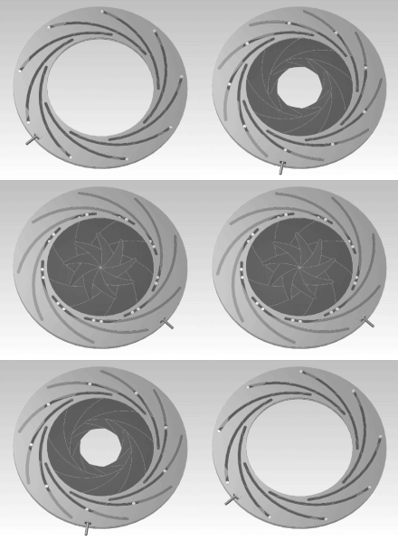
Iris diaphragm
Gap segment diaphragm
Sieve screen
Front panel, also sliding panel, push-in panel or waterhouse panel
The diaphragm is inserted / pushed into the lens through a slot. Different hole shapes are possible.
use

Photographic diaphragms are often designed as iris diaphragms ; they are adjustable aperture diaphragms for controlling the brightness and, indirectly, the depth of field of the image. A lens mount acts as a fixed aperture stop. However, since it often reflects scattered light, a fixed aperture ring is attached in front of or after it.
When enlarging negatives (working out photographs on film or plate), a right-angled snap frame made of four thin metal strips, two of which are usually adjustable, is placed over the photo paper to be exposed. This means that the paper is pressed largely flat onto the cassette (good for image sharpness and freedom from distortion), the image section is sharply defined and an unexposed, white edge is created on the paper. Contact copies of the larger glass negatives are often made in a wooden box with a fixed frame. Even the camera housing, with its film guide, mostly made of cast aluminum or plastic, creates a rectangular border of the exposed area on the film. Special features of the contour - dimensions, corner rounding, shadow of a thread of dust - provide researchers with information on the type of camera used or even the camera copy. With some films, the manufacturer not only imprints negative numbers but also separators (or frames) between the frames. Slide mounts form an optical frame around the slide film, which is typically somewhat smaller than the standard format (e.g. small picture 24 × 36 mm).
Panels with a strikingly shaped contour suggest a special perspective of the viewer in still or moving images. The classic keyhole perspective with a diaphragm made of a circle and connected trapezoid, which widens slightly towards the bottom, for an unobserved view through a door. Four squares at a short distance suggest a view through a barred prison window, many squares through a ventilation or duct inlet grille.
Small, often yellow-tinted hatches on the back of a roll film camera offer a view of the picture numbers printed on the paper strip and information on film transport and prevent light from falling on the film side. Some 35mm cameras allow this type of insight into the labeling of the film cartridge in order to be able to read off the film type.
Slit diaphragms usually restrict the beam path as field diaphragms only in one direction. In spectrometers and related optical devices, they are called optical slits , and they are usually adjustable both in terms of their width (order of magnitude 1 mm, when it comes to the visible spectral range ) and their height (order of magnitude 20 mm). They serve on the input side as secondary light sources of well-defined and easily usable form, on the output side, for example, of monochromators as selection means for certain wavelengths (ranges) and in turn as secondary light sources. The width of the gap is generally chosen so large that diffraction effects remain negligible.
In ophthalmology , a non-adjustable pinhole diaphragm (so-called stenopean gap ) is used as an aperture diaphragm for the differential diagnostic assessment of a reduction in visual acuity . The ENT doctor can see through the diaphragm of the inclined lighting mirror in the throat, nose or ear.
Aperture
The larger the aperture diaphragm, the larger the so-called aperture can be. It is the sine value of half the cone angle α of the beam emanating from an object point. Whether the aperture diaphragm is filled depends on whether the object point is illuminated at a large angle. The so-called numerical aperture is .
A related term is the relative aperture (or aperture ratio ). The diameter of the aperture diaphragm is related to the focal length of the imaging system. Example: a photo lens with a relative aperture of 1: 2 is brighter than one with 1: 3.5.
Pupil and hatch
In addition to the objective diaphragms, the terms pupil and hatch are used in geometric optics . Each aperture and each field diaphragm is initially a pupil or a hatch. The image of each is another pupil or a hatch, so that there is one entry and one exit pupil or hatch. If the objective diaphragm is located in front of the imaging system (e.g. lens) in the imaging direction, this is the entry pupil or hatch. Your image generated by the imaging device (mostly on the other side) is then the exit pupil or hatch. If the order is reversed, the assignment is reversed.
See also
literature
- Gerhard Wanner: Microscopic-botanical internship. Georg Thieme Verlag, 2004, ISBN 978-3-13-440312-1 , p. 11 ( Google book online ).
Web links
- The field diaphragm. Retrieved January 10, 2010 .
Individual evidence
- ↑ Dietrich Kühlke: Optics. Basics and Applications. 3rd, revised and expanded edition. Harri Deutsch, Frankfurt am Main 2011, ISBN 978-3-8171-1878-6 , p. 83.
- ↑ Heinz Haferkorn: Optics. Physical-technical basics and applications. 3rd, revised and expanded edition. Barth, Leipzig et al. 1994, ISBN 3-335-00363-2 , p. 277.
- ↑ Dietrich Kühlke: Optics. Basics and Applications. 3rd, revised and expanded edition. Harri Deutsch, Frankfurt am Main 2011, ISBN 978-3-8171-1878-6 , p. 88.
- ↑ Explanation for pupils cf. Dietrich Kühlke: Optics. Basics and Applications. 3rd, revised and expanded edition. Harri Deutsch, Frankfurt am Main 2011, ISBN 978-3-8171-1878-6 , p. 85.


