Uveitis

| Classification according to ICD-10 | |
|---|---|
| H20.0 | Acute and subacute iridocyclitis |
| H20.1 | Chronic iridocyclitis |
| H20.2 | Phacogenic iridocyclitis |
| H20.9 | Iridocyclitis, unspecified |
| H30.2 | Posterior cyclitis |
| H22.0 * | Iridocyclitis in Infectious and Parasitic Diseases Classified Elsewhere |
| H22.1 * | Iridocyclitis in other diseases classified elsewhere |
| ICD-10 online (WHO version 2019) | |
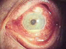
A Uveitis is an inflammation of the eyes, skin ( uvea ) selected from the choroid ( choroidal ), the beam body ( ciliary body ) and the iris ( Iris is). The vitreous humor can also be involved. If the corpus ciliare is affected, one speaks of an inflammation of the ciliary body , if the choroid is affected by an inflammation of the choroid or choroiditis .
classification
There is an abundance of different clinical pictures that can cause uveitis. One possibility for differentiation is the classification according to the anatomical location of the inflammation, i.e. according to one or more of the three parts of the uvea, a further differentiation sees the cause superficially (infectious or non-infectious, e.g. bacterial or viral):
- the anterior uveitis (in German-speaking also called iritis) is inflammation of the anterior portion of the uveal tract, particularly of the iris and the ciliary muscle. If only cells are in the anterior chamber of the eye, one speaks of iritis ; if there are also a few cells behind the lens , including in the anterior vitreous humor, one speaks of iridocyclitis . Accompanying here to macular edema and edema of the optic nerve can occur. If the inflammatory cells are arranged in a nodular manner, one speaks of a "granulomatous" inflammation.
- the intermediate uveitis concerns the central part of the uvea. Here one finds the highest density of free inflammatory cells in the vitreous body (vitreous inflammation or vitritis ). However, there can also be a few cells in the anterior chamber. A special form of intermediate uveitis is pars planitis , in which inflammatory deposits (snowdrifts, English: snowbank ) are found, especially in the lower transition area between the retina and the ciliary body. If the inflammatory cells cluster in the vitreous space , one speaks of cotton balls or snowballs . This can be accompanied by retinal periphlebitis (inflammation of the retinal veins , vasculitis ), macular edema and papillary edema.
- the posterior uveitis now also includes changes (infiltration with inflammatory cells) in the retina and choroid. Depending on the infestation, one speaks of retinitis , choroiditis and chorioretinitis or retinochoroiditis .
- the panuveitis shows in all three areas of inflammatory cells, but says nothing about the severity of inflammation.
In addition, you can use explanatory models to classify uveitis . The question arises: Can the infestation pattern on the eye be assigned to a defined disease?
A distinction is made between the following forms:
- the primary uveitis , apart from the anatomical classification (see above), cannot be further explained (approx. 40% of patients). This form was also called endogenous or idiopathic in the past
- the secondary uveitis (approximately 60% of patients), which further is divided in associated with systemic disease , infection and ocular syndrome
The masquerade forms (pseudo-uveitis) must be differentiated from both forms . These only look like this at first; however, in further diagnostics it is found that this is z. B. tumors in the eye (e.g. oculocerebral lymphoma ). The supposed inflammatory cells are tumor cells. Other pseudo-uveitids include a. the retinitis pigmentosa and the pigment dispersion syndrome.
Symptoms
This distinction is reflected in the severity of the disease. The inflammation of the posterior parts of the choroid (posterior uveitis / panuveitis) leads more often to a permanent reduction in visual acuity (acute: cloud vision, blurred vision) than an anterior uveitis, in which the reddening of the eyes is in the foreground. Symptoms such as pain, sensitivity to light, increased tearing and feelings of foreign bodies also occur. As a general rule, the further in front and outside the inflammation is anatomically located in the eye, the more discomfort it causes the patient. The typical symptoms of anterior uveitis are red eyes, pain, and sensitivity to light. The symptoms of uveitis intermedia, on the other hand, are blurred and dot vision in an externally white eye. The symptoms of posterior uveitis can either be minimal for the patient (the infiltrates are outside the point of sharpest vision) or manifest as a non-moving cloud in front of the point of sharpest vision.
The disease can - but does not have to - occur on both sides and can be mistaken for conjunctivitis by laypeople .
Certain uveitides in children with a rheumatic disease can pass without the typical uveitis symptoms and thus go unnoticed. Children with rheumatic diseases should therefore see an ophthalmologist immediately after the rheumatism is diagnosed.
Secondary forms
In contrast to the primary forms of uveitis, the secondary forms are usually characterized by typical constellations of findings. The diagnosis can often be confirmed after certain laboratory tests have been carried out or when certain general symptoms are present. The treatment of uveitis is always symptom-oriented. After the diagnosis has been confirmed, specific therapeutic procedures are carried out as far as possible, corresponding to the cause.
Different forms of uveitis are triggered by specific pathogens:
-
Viruses
- Herpes zoster ( varicella zoster virus )
- Cytomegalovirus retinitis
- Progressive external retinal necrosis ( varicella zoster virus in AIDS )
- Acute retinal necrosis ( herpes simplex virus 1 and 2, varicella zoster virus )
- Congenital rubella ( rubella virus )
- Lymphocytic choriomeningitis
-
Parasites
- Toxoplasmosis
- Toxocariasis
- Choroidal pneumocystosis
-
Mushrooms
- Histoplasmosis
- Candidiasis
- Cryptococcal choroiditis
- bacteria
The iris inflammation after infection with such germs is often not a direct eye infection. Therefore, no pathogens are found in smear tests on the eye. Iritis is often an immunological response to the body's handling of these germs, which are located elsewhere in the body. Often they do not cause symptoms there. The actual infection also precedes the iritis with a time lag.
Uveitis can be associated with certain diseases. However, these well-known associations initially say nothing about the specific cause of uveitis. Autoimmunological processes are often assumed to be the cause.
-
Spondyloarthritis
- Ankylosing spondylitis (Bechterew's disease)
- Reactive arthritis
- Psoriatic arthritis (psoriatic arthritis)
- Juvenile idiopathic arthritis
- Inflammatory bowel disease
- Kidney disease
- Non-infectious multisystem diseases
- Sarcoid (Boeck's disease)
- Behçet's disease
- Vogt-Koyanagi-Harada syndrome
Furthermore, there are certain, clinically clearly demarcated uveitis disease pictures without systemic associations, also called specific uveitis :
- Fuchs' uveitis syndrome
- (Idiopathic) intermedia uveitis
- Juvenile Chronic Iridocyclitis
- Acute anterior uveitis in adults ( HLA-B27-positive acute anterior uveitis but also HLA-B27-negative)
The (idiopathic) syndromes with multifocal white spots (English: White Dot Syndrome) are also included in the uveitids:
- Acute multifocal posterior placoid pigment epithelopathy (AMPPE)
- Serping choroidopathy
- Birdshot chorioretinopathy
- Punctiform internal choroidopathy
- Multifocal choroiditis with panuveitis
- Multiple evanescent white dot syndrome
- Acute retinal pigment epitheliitis
- MEWDS
Rheumatic disease
Irritation of the iris is a typical disease that accompanies inflammatory diseases of the spine . Inflammation of joints ( arthritis ), tendon sheaths ( tendovaginitis ) and especially of tendon attachments ( enthesopathy ) are very common. Heel pain and Achilles tendinitis are also typical , for which there is no explanation e.g. B. by an injury or an overload or overexertion.
The iris inflammation in inflammatory spinal diseases is acute, occurs suddenly, is accompanied by severe reddening of the eyes, pain and very severe visual impairment.
therapy
Treatment of uveitis depends on the severity and course. Often you get by with eye ointments containing cortisone , possibly in combination with cortisone-free anti-inflammatory drugs in the form of eye ointments or drops.
So that no sticking between the iris and lens occurs as a possible permanent consequence of the inflammation and the visual function is not permanently impaired, additional drops are given that dilate the pupil ( mydriatic ). In the case of severe iris inflammation, an injection of cortisone under the conjunctiva and / or the administration of cortisone tablets is necessary so that the eye does not permanently lose vision. In some cases, high doses of cortisone are necessary. In the case of repeated attacks, long-term therapy with low-dose corticosteroids and / or systemic immunosuppression (e.g. methotrexate, cyclosporine A, mycophenolate mofetil, etc.) is recommended.
In the event of an underlying bacterial infection, targeted antibiotic therapy is used. This must be dosed in a sufficiently high dose and carried out long enough, otherwise the pathogens will not be completely killed and relapses will occur later. The choice of antibiotics depends on the underlying germ. Antivirals are used for infections caused by viruses (e.g. Herpesviridae).
Since both the inflammatory process in the eye and the drugs used to counter it can lead to an increase in intraocular pressure, its control is important.
literature
- Marianne Abele-Horn: Antimicrobial Therapy. Decision support for the treatment and prophylaxis of infectious diseases. With the collaboration of Werner Heinz, Hartwig Klinker, Johann Schurz and August Stich, 2nd, revised and expanded edition. Peter Wiehl, Marburg 2009, ISBN 978-3-927219-14-4 , p. 115 ( Uveitis ).
- Manfred Zierhut: Intraocular Inflamation . With the collaboration of Carlos Pavesio, Shigeaki Ohno, Fernando Oréfice, Narsing A. Rao, Springer; Edition: 1st ed. 2016 (February 3, 2016) ISBN 978-3540753858