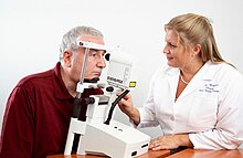
Heidelberg retina tomograph
The Heidelberg Retina Tomograph (HRT) is an ophthalmological confocal point scanning laser ophthalmoscope for examining the cornea and certain areas of the retina using different diagnostic modules (HRT Retina, HRT Cornea, HRT Glaucoma). The most widely used area of application of HRT, however, is the inspection of the optic nerve head (papilla) for the early detection and follow-up of glaucoma ( glaucoma ). In addition to the visual field examination ( perimetry ), the chamber angle examination ( gonioscopy) and intraocular pressure measurement ( tonometry ) have established themselves as an integral part of routine glaucoma diagnostics. To date, HRT has the highest resolution of all imaging methods for diagnosing glaucoma. In Germany , its use represents an individual health service ( IGeL ), the costs of which have not yet been covered by health insurance companies .
Glaucoma diagnostics
During the examination, a laser beam passes through the pupil opening onto the fundus and scans the optic nerve head ( papilla ) and the retina . A three-dimensional height relief is generated from tens of thousands of measuring points, which allows a quantitative assessment of all relevant anatomical structures:
- Papilla excavation (shape, asymmetry),
- neuroretinal rim (area and volume) and
- peripapillary retinal nerve fiber layer (height variation of the retinal surface, thickness, asymmetry).
These stereometric parameters are compared with extensive databases and thus enable a classification of the eye taking into account the individual papilla size and patient age. Two independent classification procedures based on different approaches are available.
Moorfields Regression Analysis (MRA)
This method takes physiological relationships into account, for example the dependence of the size of the neuroretinal rim on the size of the papilla, and the decrease in the size of the rim with increasing age. The MRA classifies an eye as being inside or outside normal limits. The classification result is given separately for the entire papilla and for six individual sectors.
Glaucoma Probability Analysis (GPS)
The shape of the optic nerve head changes as the glaucoma progresses. The system uses artificial intelligence methods to classify an eye based on the shape of the papilla and the peripapillary retinal nerve fiber layer. The following structures are included in the model:
- Excavation (excavation size, excavation depth, steepness of the edge),
- Retinal nerve fiber layer (horizontal curvature of the peripapillary nerve fiber layer, vertical curvature of the peripapillary nerve fiber layer).
This model from all essential anatomical structures is compared with an extensive database of normal and early glaucomatous eyes. The GPS classification method is considered objective and user-independent.
Topographical Change Analysis (TCA)
If glaucoma is suspected, the occurrence of progression (progressive degeneration of the optic nerve) decides the diagnosis. In the case of manifest glaucoma, the rate of progression is an important measure for the treatment decision. Glaucoma experts from the Association of International Glaucoma Societies (AIGS) have therefore proposed “progressive structural changes in the optic nerve head” as an assessment standard in glaucoma diagnostics. With the appropriate module, the HRT is able to meet this requirement and ensure the precise analysis of changes over time within the three anatomical structures that are important for glaucoma during follow-up checks. The determination of significant and reproducible changes through extensive data analyzes and comparisons has been proven in various long-term studies.
Glaucoma early detection
The Ocular Hypertension Treatment Study (OHTS) has demonstrated that imaging tests can detect the onset of glaucoma before visual field or clinical disc evaluation reveals any detectable abnormalities. In this study, patients with high intraocular pressure but normal visual field and, according to expert findings, normal structure of the optic nerve head were examined. It was shown that a positive result with the HRT had the highest predictive value for the later development of glaucoma. The onset of structural glaucomatous changes in the papilla was detected up to eight years before a positive visual field finding and before a papillary change that could be recognized by glaucoma experts on the basis of stereo fundus photos. Studies with HRT allow patients with increased intraocular pressure to be divided into high and low risk groups.
Retinal diagnostics
A special diagnostic module enables detailed examinations of the retina to be carried out using three-dimensional images. On the one hand, using the so-called edema index, areas are made visible that have increased fluid retention. On the other hand, the HRT is able to measure the retinal thickness. The main area of application is therefore the examination of macular and retinal edema.
Corneal diagnostics
With the use of special additional optics, the examiner can display corneal layers of different depths with the HRT and thus make the finest cells visible in every layer. This allows for a relatively early clinical assessment of various corneal diseases.
literature
- Friedrich E. Kruse, Reinhard O. Burk, Hans E. Völcker, Gerhard Zinser, Ulrich Harbarth: Reproducibility of topographic measurements of the optic nerve head with laser tomographic scanning . In: Ophthalmology . tape 96 , no. 9 , 1989, ISSN 1549-4713 , pp. 1320-1324 , doi : 10.1016 / S0161-6420 (89) 32719-9 , PMID 2780001 (first description).
- Reinhard OW Burk, Klaus Rohrschneider, Friedrich E. Kruse, Hans E. Völcker: Laser scanning tomography of the papilla . In: Eugen Gramer (Ed.): Glaucoma. Diagnostics and therapy . Enke, Stuttgart 1990, ISBN 3-432-98691-2 , pp. 113-119 .
- P. Janknecht, J. Funk: Optic nerve head analyzer and Heidelberg retina tomograph: accuracy and reproducibility of topographic measurements in a model eye and in volunteers . In: British Journal of Ophthalmology . tape 78 , no. 10 , 1994, ISSN 0007-1161 , pp. 760-768 , doi : 10.1136 / bjo.78.10.760 , PMID 7803352 .
- Ville Saarela, P. Juhani Airaksinen: Heidelberg retina tomograph parameters of the optic disc in eyes with progressive retinal nerve fiber layer defects . In: Acta Ophthalmologica . tape 86 , no. 6 , 2008, ISSN 1755-375X , p. 603-608 , doi : 10.1111 / j.1600-0420.2007.01119.x , PMID 18752515 .
- M. Durmus, R. Karadag, M. Erdurmus, Y. Totan, I. Feyzi Hepsen: Assessment of cup-to-disc ratio with slit-lamp funduscopy, Heidelberg Retina Tomography II, and stereoscopic photos . In: European Journal of Ophthalmology . tape 19 , no. 1 , 2009, ISSN 1120-6721 , p. 55-60 , PMID 19123149 .
- Albert J. Augustin: Ophthalmology. 3rd, completely revised and expanded edition. Springer, Berlin et al. 2007, ISBN 978-3-540-30454-8 , p. 961 ff.
Web links
Individual evidence
- ↑ Marc Makert: Confocal in-vivo microscopy of the conjunctiva (PDF; 9.5 MB)
- ↑ AAD - Ophthalmological Academy of Germany ( Memento of the original from December 16, 2013 in the Internet Archive ) Info: The archive link was inserted automatically and not yet checked. Please check the original and archive link according to the instructions and then remove this notice.
- ↑ Gad Wollstein, David F. Garway-Heath, Roger A. Hitchings: Identification of early glaucoma cases with the scanning laser ophthalmoscope11 The authors have no proprietary interest in the development or marketing of this or a competing instrument. In: Ophthalmology . tape 105 , no. 8 , 1998, pp. 1557-1563 , doi : 10.1016 / S0161-6420 (98) 98047-2 .
- ↑ David F. Garway-Heath: Moorfields Regression Analysis . In: Murray Fingeret, John G. Flanagan, Jeffrey M. Liebmann (Eds.): HRT Fibel . Engineering, Heidelberg 2006, p. 31–39 ( PDF; 210 MB ). PDF; 210 MB ( Memento of the original from January 6, 2011 in the Internet Archive ) Info: The archive link was inserted automatically and has not yet been checked. Please check the original and archive link according to the instructions and then remove this notice.
- ↑ Nicholas V. Swindale, Gordana Stjepanovic, Adeline Chin, Frederick S. Mikelberg: Automated Analysis of Normal and Glaucomatous Optic Nerve Head Topography Images . In: Investigative Ophthalmology & Visual Science . tape 41 , no. 7 , 2000, ISSN 0146-0404 , p. 1730-1742 .
- ^ Robert N. Weinreb, Erik L. Greve (Eds.): Glaucoma Diagnosis. Structure and Function. Reports and Consensus Statements of the 1st Global AIGS Consensus Meeting on "Structure and function in the management of glaucoma" (= Consensus Series. Vol. 1). Kugler Publications, The Hague 2004, ISBN 90-6299-200-5 .
- ↑ Balwantray C. Chauhan, Terry A. McCormick, Marcelo T. Nicolela, Raymond P. LeBlanc: Optic Disc and Visual Field Changes in a Prospective Longitudinal Study of Patients With GlaucomaComparison of Scanning Laser Tomography With Conventional Perimetry and Optic Disc Photography . In: Archives of Ophthalmology . tape 119 , no. 10 , 2001, ISSN 0003-9950 , p. 1492–1499 , doi : 10.1001 / archopht.119.10.1492 .
- ↑ DS Kamal, DF Garway-Heath, RA Hitchings, FW Fitzke: Use of sequential Heidelberg retina tomograph images to identify changes at the optic disc in ocular hypertensive patients at risk of developing glaucoma . In: British Journal of Ophthalmology . tape 84 , no. 9 , 2000, ISSN 0007-1161 , p. 993-998 , doi : 10.1136 / bjo.84.9.993 .
- ↑ DF Garway-Heath, G. Wollstein, RA Hitchings: Aging changes of the optic nerve head in relation to open angle glaucoma . In: British Journal of Ophthalmology . tape 81 , no. 10 , January 10, 1997, p. 840-845 , doi : 10.1136 / bjo.81.10.840 .
- ↑ Christopher Bowd, Linda M. Zangwill, Felipe A. Medeiros, Jiucang Hao, Kwokleung Chan, Te-Won Lee, Terrence J. Sejnowski, Michael H. Goldbaum, Pamela A. Sample, Jonathan G. Crowston, Robert N. Weinreb: Confocal Scanning Laser Ophthalmoscopy Classifiers and Stereophotograph Evaluation for Prediction of Visual Field Abnormalities in Glaucoma-Suspect Eyes . In: Investigative Ophthalmology & Visual Science . tape 45 , no. 7 , 2004, ISSN 0146-0404 , p. 2255-2262 , doi : 10.1167 / iovs.03-1087 .
- ↑ Gadi Wollstein, David F. Garway-Heath, Darmalingum Poinoosawmy, Roger A. Hitchings: Glaucomatous optic disc changes in the contralateral eye of unilateral normal pressure glaucoma patients . In: Ophthalmology . tape 107 , no. 12 , 2000, pp. 2267-2271 .
- ↑ Christopher Bowd, Madhusudhanan Balasubramanian, Robert N. Weinreb, Gianmarco Vizzeri, Luciana M. Alencar, Neil O'Leary, Pamela A. Sample, Linda M. Zangwill: Performance of Confocal Scanning Laser Tomograph Topographic Change Analysis (TCA) for Assessing Glaucomatous Progression . In: Investigative Ophthalmology & Visual Science . tape 50 , no. 2 , 2009, p. 691-701 , doi : 10.1167 / iovs.08-2136 .
- ↑ Linda M. Zangwill, Robert N. Weinreb, Julia A. Beiser, Charles C. Berry, George A. Cioffi, Anne L. Coleman, Gary Trick, Jeffrey M. Liebmann, James D. Brandt, Jody R. Piltz-Seymour , Keri A. Dirkes, Suzanne Vega, Michael A. Kass, Mae O. Gordon: Baseline Topographic Optic Disc Measurements Are Associated With the Development of Primary Open-Angle Glaucoma The Confocal Scanning Laser Ophthalmoscopy Ancillary Study to the Ocular Hypertension Treatment Study . In: Archives of Ophthalmology . tape 123 , no. 9 , September 1, 2005, pp. 1188–1197 , doi : 10.1001 / archopht.123.9.1188 .


