Pillar bell
| Pillar bell | ||||||||||||
|---|---|---|---|---|---|---|---|---|---|---|---|---|

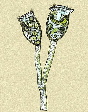
Pillar bells ( Epistylis sp. , Drawing after a microscope view) |
||||||||||||
| Systematics | ||||||||||||
|
||||||||||||
| Scientific name | ||||||||||||
| Epistyle | ||||||||||||
| Ehrenberg , 1830 |
Column bells ( epistylis ) form a genus of unicellular ciliate animals . Like the other species of the Epistylidae family , the columnar bells live sessile on aquatic plants and detritus, but also as ectocommensals on the body surface of crustaceans , fish and turtles . They attach themselves to the surface of the animals by means of a stalk and use their eyelashes to swirl food into their inverted bell-shaped cell bodies.
features
The individual cells can be 10 to 250 micrometers long and 10 to 80 micrometers wide, depending on the type. Their stems are branched and form colonies. At the end of each stem there is an individual (zooid). The colonies reach a length of several millimeters and are macroscopically visible as growth. There is no muoneme inside the stalk as in the bell animals , so that the colonies cannot contract as in Vorticella or Zoothamnium .
The stem is dichotomously branched, after each fork one stem is shorter than the other. A separation zone is created on the shorter one before each division. At these points, the oldest branches can be shed and float freely through the water until they find a suitable place to form a new colony.
Further features of the genus are a broad lip formation at the apical end and a continuous, simple lash line around the mouth. In related genera such as Campanella or Heteropolaria , this spiral has one and a half or four to six whorls. Opercularia and Orbopercularia have no lip formation.
Way of life
In contrast to the ectoparasites , ectocommensals are mostly harmless. They live on the food waste from their host animals or, like Epistylis, mainly on bacteria that decompose the waste.
Mass infestation
In environments where the nutrient content in the water is very high due to pollution, epistylis can multiply on a mass scale . This can cause skin irritation and ulcers in the fish, making them susceptible to infection.
Fish disease
An excessive infestation of fish by Epistylis be determined by small white, cotton-like growths on the size of a grain of rice. The infestation can occur on any part of the body, but it is usually first visible on the side lines of the fish. In places with of mass infection with Epistylis unicellular often occur bloody wounds by secretions. Mass infestation of the gills can affect the breathing of the fish.
An initial diagnosis can be made with the naked eye, but a more precise determination can only be made with a microscope.
Infestation by crayfish
Also in crayfish the massive infestation is Epistylis an indicator of polluted water and low oxygen concentration (high BOD5 value of the oxygen consumption). The infestation can affect the quality of production in cancer farms. Increased epistyle growth takes place at higher water temperatures. Here, too, a macroscopically visible white or gray fluff forms on the exoskeleton of the crabs.
Prevention and treatment
Clean, not too warm water is the best prerequisite for preventing mass infestation. Appropriate feeding prevents too much organic material in the pond or aquarium. Lower stocking density helps to avoid contagion. In the aquarium, a filter with UV light can kill the epistyle and other pathogens.
The use of iodine-free sodium chloride (NaCl) in a concentration of up to 0.6% for a maximum of 10 days or a mixture of formaldehyde and salt in the water achieve the best effect.
Systematics
The genus Epistylis is unlikely to be monophyletic , that is, not all species are descended from a common ancestor. Epistylis galea , for example, shows a polymorphism of the individual individuals within a colony. As with many Zoothamnium species, macro and micro zooids occur. Although Fauré-Fremiet already suspected in 1907 that Epistylis galea could be assigned to the genus Campanella , only molecular genetic studies at the Institute of Microbiology of the Chinese Academy of Sciences in 2005 showed a greater relationship between Epistylis galea and Campanella - and Opercularia species than other Epistylis - Species. The molecular genetic investigation of 18S-rRNA sequences of various sessile ciliates from the subclass Peritrichia also showed the result that the genera Epistylis and the bell animals (genus Vorticella ) cannot be monophyletic. It is assumed that the morphological features that were previously used to classify the Peritrichia are insufficient for a phylogenetic system. A revision of the taxa is not possible until no further characteristics are found according to which the individual groups can be distinguished from one another.
Individual evidence
- ↑ Description and species list ( Memento of the original dated February 27, 2008 in the Internet Archive ) Info: The archive link was inserted automatically and not yet checked. Please check the original and archive link according to the instructions and then remove this notice. (engl.)
- ^ RE Klinger & RF Floyd: Red Sore Disease in game fish. Fact Sheet VM 85, Institute of Food and Agricultural Science, University of Florida, 2002
- ^ Government of Western Australia. Department of Fisheries (English; PDF; 156 kB)
- ^ WA Hubert, MC Warner: Control of Epistylis on channel catfish in raceways. In: Journal of wildlife diseases. Volume 11, Number 2, April 1975, pp. 241-244, PMID 806710 .
- ^ E. Fauré-Fremiet: L'Epistylis galea. Compt. Rend. Soc. Biol., 62, pp. 1058, 1907
- ↑ Wei Miao, Wei-Song Fen, Yu-He Yu, Xi-Yuan Zhang and Yun-Fen Shen: Phylogenetic Relationships of the Subclass Peritrichia (Oligohymenophorea, Ciliophora) Inferred from Small Subunit rRNA Gene Sequences . Journal of Eukaryotic Microbiology, 51 (2), pp. 180-186, July 2005. PMID 15134253 . doi : 10.1111 / j.1550-7408.2004.tb00543.x
- ↑ Laura RP Utz and Eduardo Eizirik: Molecular Phylogenetics of Subclass Peritrichia (Ciliophora: Oligohymenophorea) Based on Expanded Analyzes of 18S rRNA Sequence . Journal of Eukaryotic Microbiology, 54 (3), pp. 303-305, May 2007. PMID 17552986 . doi : 10.1111 / j.1550-7408.2007.00260.x
literature
- Colin R. Curds, Michael A. Gates, and David McL. Roberts: British and other freshwater ciliated protozoa Part II Ciliophora: Oligohymenophora and Polyhymenophora. Cambridge University Press, 1983
