Fontanel
The term fontanelle ( old French: "small source") is mainly used today as a medical-anatomical term. Fontanelle refers to the area of the skull of newborns or infants or more generally of newborn vertebrates that is not yet encompassed by bony or cartilaginous structures . It is a continuous unit of inner and outer membranous layers on the skull in places without bony or cartilaginous layers. There are places where at least three cover plates of the skull are not yet completely adjacent to one another.
anatomy
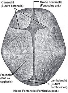
The newborn person has two unpaired main fontanels, which are referred to as large fontanels and small fontanels, and four other, smaller fontanels (paired front and rear side fontanels).
The large, diamond-shaped fontanel (forehead fontanel or fonticulus anterior) is located centrally on the skull. At this point meet each the right and left front legs (bones frontalia) and the parietal bones (parietal bones) or coronal suture (coronal suture), the sagittal suture (sagittal suture) and end seam (frontal suture) to each other (see also suture ).
The triangular small fontanelle (occipital fontanelle or fonticulus posterior) is located on the back of the head , at the point of contact of the parietal bones with the occipital bone (os occipitale) or the arrow suture with the lambda suture (sutura lambdoidea).
The posterior lateral fontanelles (Fonticulus mastoideus) lie on both sides of the head between the temporal , parietal and occipital bones.
The anterior lateral fontanelles (Fonticulus sphenoidalis) are also located on both sides of the head, between the temporal, frontal and parietal bones and the large wing of the sphenoid bone .
The fontanelles play an important role , especially during birth : Together with the cartilaginous lining of the skull, they enable the skull to be deformed during the birth by sliding the bone plates over one another , thus making it easier to pass through the birth canal. The final closure takes place after about two months for the small fontanel and after two years for the large fontanel. The side fountains close after about a year. In the case of various deficiency symptoms , such as rickets , the closure takes significantly longer. The premature ossification is called craniosynostosis (or premature suture synostosis ). Due to the soft bone plates, one-sided positioning of the infant can lead to an asymmetrically deformed skull (or plagiocephaly ) or a flat back of the head (or brachycephaly ) in the first months of life , whereby the plagiocephalus must be treated therapeutically.
In some children with Down syndrome (trisomy 21), another opening appears on the suture ( sutura sagittalis ) between the large and small fontanel, which is also known as the third fontanel .
Brachycephalic dog breeds (especially Chihuahua and Yorkshire Terriers ) tend to have fontanelles that persist throughout their lives in the area of the skull.
literature
- W. Schuster, D. Färber (editor): Children's radiology. Imaging diagnostics. Springer 1996, Vol. I, S 383ff, ISBN 3-540-60224-0 .
- E. Richter, W. Lierse: Radiological anatomy of the newborn for x-rays, sonography, CT, MRI, 1990 Urban & Schwarzenberg, ISBN 3-541-13141-1
Web links
Individual evidence
- ^ Willibald Pschyrembel : Clinical Dictionary , 266th, updated edition, de Gruyter, Berlin 2014, ISBN 978-3-11-033997-0, keyword fontanelle
- ↑ Entry on Fontanelle in Flexikon , a Wiki of the DocCheck company
- ↑ K. Stoevesandt, H. Ma, U. Beyer, H. Zhang, G. Jorch: Lagerungsplagiocephalus beim Säugling In: Monthly Pediatric Medicine (2018) Vol. 166, No. 8, pp. 675-682. (last accessed on November 14, 2018).

